 Movie
Movie Controller

[English] 日本語
 Yorodumi
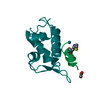
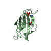
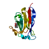
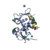
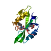
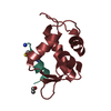
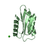
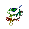
- PDB-5ema: Crystal structure of the SNX27 PDZ domain bound to the phosphoryl... -
+ Open data

- Basic information
| Entry | Database: PDB / ID: 5ema | |||||||||
|---|---|---|---|---|---|---|---|---|---|---|
| Title | Crystal structure of the SNX27 PDZ domain bound to the phosphorylated C-terminal LRRC3B PDZ binding motif | |||||||||
 Components Components |
| |||||||||
 Keywords Keywords | PROTEIN TRANSPORT / Endosome / PDZ domain / sorting nexin | |||||||||
| Function / homology |  Function and homology information Function and homology informationpostsynaptic early endosome / establishment of protein localization to plasma membrane / positive regulation of AMPA glutamate receptor clustering / neurotransmitter receptor transport to plasma membrane / postsynaptic recycling endosome / WASH complex / retromer complex / response to methamphetamine hydrochloride / endosome to plasma membrane protein transport / phosphatidylinositol-3-phosphate binding ...postsynaptic early endosome / establishment of protein localization to plasma membrane / positive regulation of AMPA glutamate receptor clustering / neurotransmitter receptor transport to plasma membrane / postsynaptic recycling endosome / WASH complex / retromer complex / response to methamphetamine hydrochloride / endosome to plasma membrane protein transport / phosphatidylinositol-3-phosphate binding / endocytic recycling / regulation of synapse maturation / endosomal transport / endosome to lysosome transport / immunological synapse / regulation of postsynaptic membrane neurotransmitter receptor levels / phosphatidylinositol binding / ionotropic glutamate receptor binding / cellular response to nerve growth factor stimulus / intracellular protein transport / Schaffer collateral - CA1 synapse / calcium-dependent protein binding / nervous system development / extracellular matrix / early endosome membrane / early endosome / endosome / postsynapse / glutamatergic synapse / signal transduction / : / membrane / cytosol Similarity search - Function | |||||||||
| Biological species |   Homo sapiens (human) Homo sapiens (human) | |||||||||
| Method |  X-RAY DIFFRACTION / SYNCHROTRON / MOLECULAR REPLACEMENT / Resolution: 1.32 Å X-RAY DIFFRACTION / SYNCHROTRON / MOLECULAR REPLACEMENT / Resolution: 1.32 Å | |||||||||
 Authors Authors | Collins, B.M. / Clairfeuille, T. | |||||||||
| Funding support |  Australia, 2items Australia, 2items
| |||||||||
 Citation Citation | Journal: Nat.Struct.Mol.Biol. / Year: 2016 Title: A molecular code for endosomal recycling of phosphorylated cargos by the SNX27-retromer complex. Authors: Clairfeuille, T. / Mas, C. / Chan, A.S. / Yang, Z. / Tello-Lafoz, M. / Chandra, M. / Widagdo, J. / Kerr, M.C. / Paul, B. / Merida, I. / Teasdale, R.D. / Pavlos, N.J. / Anggono, V. / Collins, B.M. | |||||||||
| History |
|
- Structure visualization
| Structure viewer | Molecule: MolmilJmol/JSmol |
|---|
- Downloads & links
-Download
| PDBx/mmCIF format | 5ema.cif.gz | 55.5 KB | Display | PDBx/mmCIF format |
|---|---|---|---|---|
| PDB format | pdb5ema.ent.gz | 39.1 KB | Display | PDB format |
| PDBx/mmJSON format | 5ema.json.gz | Tree view | PDBx/mmJSON format | |
| Others |  Other downloads Other downloads |
-Validation report
| Arichive directory | https://data.pdbj.org/pub/pdb/validation_reports/em/5emaftp://data.pdbj.org/pub/pdb/validation_reports/em/5ema | HTTPS FTP |
|---|
-Related structure data
| Related structure data | 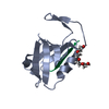 5elqC  5em9C  5embC  4z8jS S: Starting model for refinement C: citing same article ( |
|---|---|
| Similar structure data |


-Links
 PDBj
PDBj


- Assembly
| Deposited unit | 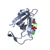
| ||||||||
|---|---|---|---|---|---|---|---|---|---|
| 1 |
| ||||||||
| Unit cell |
|
-Components
| #1: Protein | Mass: 10569.952 Da / Num. of mol.: 1 / Fragment: PDZ domain (UNP residues 39-133) Source method: isolated from a genetically manipulated source Source: (gene. exp.)  |
|---|---|
| #2: Protein/peptide | Mass: 924.886 Da / Num. of mol.: 1 / Source method: obtained synthetically / Source: (synth.) Homo sapiens (human) / References: UniProt: Q96PB8*PLUS |
| #3: Chemical | ChemComp-GOL /   Mass: 92.094 Da / Num. of mol.: 1 / Source method: obtained synthetically / Formula: C3H8O3 Mass: 92.094 Da / Num. of mol.: 1 / Source method: obtained synthetically / Formula: C3H8O3 |
| #4: Water | ChemComp-HOH /  Mass: 18.015 Da / Num. of mol.: 150 / Source method: isolated from a natural source / Formula: H2O Mass: 18.015 Da / Num. of mol.: 150 / Source method: isolated from a natural source / Formula: H2O |
| Has protein modification | Y |
-Experimental details
-Experiment
| Experiment | Method: X-RAY DIFFRACTION / Number of used crystals: 1 |
|---|
- Sample preparation
| Crystal | Density Matthews: 2.2 Å3/Da / Density % sol: 44.2 % |
|---|---|
| Crystal grow | Temperature: 293 K / Method: vapor diffusion / pH: 4.6 Details: 2 M ammonium sulphate and 0.1 M sodium acetate (pH 4.6) PH range: 4.6 |
-Data collection
| Diffraction | Mean temperature: 100 K |
|---|---|
| Diffraction source | Source: SYNCHROTRON / Site: Australian Synchrotron / Beamline: MX2 / Wavelength: 0.9537 Å |
| Detector | Type: ADSC QUANTUM 210r / Detector: CCD / Date: Oct 1, 2015 |
| Radiation | Protocol: SINGLE WAVELENGTH / Monochromatic (M) / Laue (L): M / Scattering type: x-ray |
| Radiation wavelength | Wavelength: 0.9537 Å / Relative weight: 1 |
| Reflection | Resolution: 1.32→48.5 Å / Num. obs: 23850 / % possible obs: 96.7 % / Redundancy: 7.1 % / Rmerge(I) obs: 0.054 / Net I/σ(I): 14.9 |
| Reflection shell | Resolution: 1.32→1.34 Å / Redundancy: 6.2 % / Rmerge(I) obs: 0.614 / Mean I/σ(I) obs: 2.5 / % possible all: 86.4 |
- Processing
| Software |
| |||||||||||||||||||||||||||||||||||||||||||||||||||||||||||||||||||||||||||||||||||||||||||||||||||||||||
|---|---|---|---|---|---|---|---|---|---|---|---|---|---|---|---|---|---|---|---|---|---|---|---|---|---|---|---|---|---|---|---|---|---|---|---|---|---|---|---|---|---|---|---|---|---|---|---|---|---|---|---|---|---|---|---|---|---|---|---|---|---|---|---|---|---|---|---|---|---|---|---|---|---|---|---|---|---|---|---|---|---|---|---|---|---|---|---|---|---|---|---|---|---|---|---|---|---|---|---|---|---|---|---|---|---|---|
| Refinement | Method to determine structure: MOLECULAR REPLACEMENT Starting model: 4Z8J Resolution: 1.32→36.721 Å / SU ML: 0.11 / Cross valid method: FREE R-VALUE / σ(F): 1.36 / Phase error: 16.8 / Stereochemistry target values: ML
| |||||||||||||||||||||||||||||||||||||||||||||||||||||||||||||||||||||||||||||||||||||||||||||||||||||||||
| Solvent computation | Shrinkage radii: 0.9 Å / VDW probe radii: 1.11 Å / Solvent model: FLAT BULK SOLVENT MODEL | |||||||||||||||||||||||||||||||||||||||||||||||||||||||||||||||||||||||||||||||||||||||||||||||||||||||||
| Refinement step | Cycle: LAST / Resolution: 1.32→36.721 Å
| |||||||||||||||||||||||||||||||||||||||||||||||||||||||||||||||||||||||||||||||||||||||||||||||||||||||||
| Refine LS restraints |
| |||||||||||||||||||||||||||||||||||||||||||||||||||||||||||||||||||||||||||||||||||||||||||||||||||||||||
| LS refinement shell |
|
