cellular response to vitamin B1 / response to formaldehyde / response to water-immersion restraint stress / response to ether / traversing start control point of mitotic cell cycle / atrial septum development / fibroblast activation / regulation of protein catabolic process at postsynapse, modulating synaptic transmission / Trafficking of AMPA receptors / negative regulation of intrinsic apoptotic signaling pathway by p53 class mediator ...cellular response to vitamin B1 / response to formaldehyde / response to water-immersion restraint stress / response to ether / traversing start control point of mitotic cell cycle / atrial septum development / fibroblast activation / regulation of protein catabolic process at postsynapse, modulating synaptic transmission / Trafficking of AMPA receptors / negative regulation of intrinsic apoptotic signaling pathway by p53 class mediator / receptor serine/threonine kinase binding / negative regulation of protein processing / response to steroid hormone / SUMO transferase activity / peroxisome proliferator activated receptor binding / positive regulation of vascular associated smooth muscle cell migration / atrioventricular valve morphogenesis / ventricular septum development / AKT phosphorylates targets in the cytosol / response to iron ion / NEDD8 ligase activity / endocardial cushion morphogenesis / cellular response to peptide hormone stimulus / positive regulation of muscle cell differentiation / cardiac septum morphogenesis / regulation of postsynaptic neurotransmitter receptor internalization / SUMOylation of ubiquitinylation proteins / blood vessel development / cellular response to alkaloid / ligase activity / Constitutive Signaling by AKT1 E17K in Cancer / negative regulation of DNA damage response, signal transduction by p53 class mediator / cellular response to antibiotic / SUMOylation of transcription factors / negative regulation of signal transduction by p53 class mediator / regulation of protein catabolic process / cellular response to UV-C / cellular response to estrogen stimulus / response to magnesium ion / protein sumoylation / blood vessel remodeling / ribonucleoprotein complex binding / protein localization to nucleus / positive regulation of vascular associated smooth muscle cell proliferation / protein autoubiquitination / positive regulation of mitotic cell cycle / centriolar satellite / NPAS4 regulates expression of target genes / transcription repressor complex / regulation of heart rate / : / positive regulation of protein export from nucleus / DNA damage response, signal transduction by p53 class mediator / response to cocaine / sperm end piece / ubiquitin binding / establishment of protein localization / sperm principal piece / Stabilization of p53 / cellular response to gamma radiation / cellular response to growth factor stimulus / Regulation of RUNX3 expression and activity / RING-type E3 ubiquitin transferase / Oncogene Induced Senescence / Regulation of TP53 Activity through Methylation / protein destabilization / Degradation of CDH1 / response to toxic substance / cellular response to hydrogen peroxide / disordered domain specific binding / protein polyubiquitination / p53 binding / ubiquitin-protein transferase activity / endocytic vesicle membrane / Signaling by ALK fusions and activated point mutants / Regulation of TP53 Degradation / ubiquitin protein ligase activity / negative regulation of neuron projection development / positive regulation of proteasomal ubiquitin-dependent protein catabolic process / sperm midpiece / protein-containing complex assembly / 5S rRNA binding / Oxidative Stress Induced Senescence / cellular response to hypoxia / Regulation of TP53 Activity through Phosphorylation / amyloid fibril formation / ubiquitin-dependent protein catabolic process / proteasome-mediated ubiquitin-dependent protein catabolic process / regulation of cell cycle / postsynaptic density / Ub-specific processing proteases / response to xenobiotic stimulus / protein ubiquitination / protein domain specific binding / response to antibiotic / negative regulation of DNA-templated transcription / apoptotic process / positive regulation of cell population proliferation / positive regulation of gene expression / ubiquitin protein ligase binding Similarity search - Function
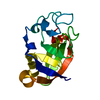
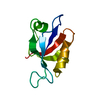
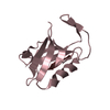
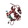
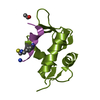
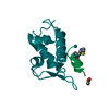
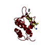
E3 ubiquitin-protein ligase Mdm2 / MDM2, modified RING finger, HC subclass / MDM2 / SWIB/MDM2 domain / p53 negative regulator Mdm2/Mdm4 / SWIB/MDM2 domain / SWIB/MDM2 domain / SWIB/MDM2 domain profile. / SWIB/MDM2 domain superfamily / Zn-finger in Ran binding protein and others ...E3 ubiquitin-protein ligase Mdm2 / MDM2, modified RING finger, HC subclass / MDM2 / SWIB/MDM2 domain / p53 negative regulator Mdm2/Mdm4 / SWIB/MDM2 domain / SWIB/MDM2 domain / SWIB/MDM2 domain profile. / SWIB/MDM2 domain superfamily / Zn-finger in Ran binding protein and others / Zinc finger, C3HC4 type (RING finger) / Zinc finger RanBP2 type profile. / Zinc finger, RanBP2-type superfamily / Zinc finger RanBP2-type signature. / Zinc finger, RanBP2-type / Zinc finger RING-type profile. / Zinc finger, RING-type / Zinc finger, RING/FYVE/PHD-type / Orthogonal Bundle / Mainly Alpha Similarity search - Domain/homology
Resolution: 1.8→41.7 Å / Cor.coef. Fo:Fc: 0.94 / Cor.coef. Fo:Fc free: 0.924 / SU B: 3.755 / SU ML: 0.079 / Cross valid method: THROUGHOUT / ESU R: 0.028 / ESU R Free: 0.029 / Stereochemistry target values: MAXIMUM LIKELIHOOD / Details: HYDROGENS HAVE BEEN ADDED IN THE RIDING POSITIONS
Rfactor
Num. reflection
% reflection
Selection details
Rfree
0.24656
6374
5.2 %
RANDOM
Rwork
0.19793
-
-
-
obs
0.20046
116968
95.21 %
-
Solvent computation
Ion probe radii: 0.8 Å / Shrinkage radii: 0.8 Å / VDW probe radii: 1.2 Å / Solvent model: MASK
 Movie
Movie Controller

 Yorodumi
 Open data
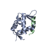
 Basic information
 Components
 Keywords
 Function and homology information
 Homo sapiens (human)
 X-RAY DIFFRACTION /
  Authors
 United States, 1items
  Citation
 Structure visualization
 Downloads & links
 Other downloads


 PDBj
PDBj


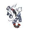
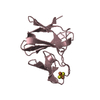
 Assembly
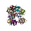


 Mass: 35.453 Da / Num. of mol.: 2 / Source method: obtained synthetically / Formula: Cl
 Mass: 18.015 Da / Num. of mol.: 418 / Source method: isolated from a natural source / Formula: H2O
 Sample preparation
 Processing