+ Open data
- Basic information
| Entry | Database: PDB / ID: 3hfn | ||||||
|---|---|---|---|---|---|---|---|
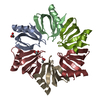
| Title | Crystal Structure of an Hfq protein from Anabaena sp. | ||||||
 Components Components | Asl2047 protein | ||||||
 Keywords Keywords | RNA BINDING PROTEIN / HFQ / SM / RNA-BINDING PROTEIN / SRNA / TRANSLATIONAL REGULATION | ||||||
| Function / homology |  Function and homology information Function and homology information | ||||||
| Biological species |  Nostoc sp. (bacteria) Nostoc sp. (bacteria) | ||||||
| Method |  X-RAY DIFFRACTION / X-RAY DIFFRACTION /  SYNCHROTRON / SYNCHROTRON /  MOLECULAR REPLACEMENT / MOLECULAR REPLACEMENT /  molecular replacement / Resolution: 2.31 Å molecular replacement / Resolution: 2.31 Å | ||||||
 Authors Authors | Boggild, A. / Overgaard, M. / Valentin-Hansen, P. / Brodersen, D.E. | ||||||
 Citation Citation |  Journal: Febs J. / Year: 2009 Journal: Febs J. / Year: 2009Title: Cyanobacteria contain a structural homologue of the Hfq protein with altered RNA-binding properties. Authors: Boggild, A. / Overgaard, M. / Valentin-Hansen, P. / Brodersen, D.E. | ||||||
| History |
|
- Structure visualization
| Structure viewer | Molecule:  Molmil Molmil Jmol/JSmol Jmol/JSmol |
|---|
- Downloads & links
- Download
| PDBx/mmCIF format |  3hfn.cif.gz 3hfn.cif.gz | 35.8 KB | Display |  PDBx/mmCIF format PDBx/mmCIF format |
|---|---|---|---|---|
| PDB format |  pdb3hfn.ent.gz pdb3hfn.ent.gz | 24.3 KB | Display |  PDB format PDB format |
| PDBx/mmJSON format |  3hfn.json.gz 3hfn.json.gz | Tree view |  PDBx/mmJSON format PDBx/mmJSON format | |
| Others |  Other downloads Other downloads |
-Validation report
| Summary document |  3hfn_validation.pdf.gz 3hfn_validation.pdf.gz | 434.3 KB | Display |  wwPDB validaton report wwPDB validaton report |
|---|---|---|---|---|
| Full document |  3hfn_full_validation.pdf.gz 3hfn_full_validation.pdf.gz | 443.3 KB | Display | |
| Data in XML |  3hfn_validation.xml.gz 3hfn_validation.xml.gz | 7.8 KB | Display | |
| Data in CIF |  3hfn_validation.cif.gz 3hfn_validation.cif.gz | 9.4 KB | Display | |
| Arichive directory |  https://data.pdbj.org/pub/pdb/validation_reports/hf/3hfn https://data.pdbj.org/pub/pdb/validation_reports/hf/3hfn ftp://data.pdbj.org/pub/pdb/validation_reports/hf/3hfn ftp://data.pdbj.org/pub/pdb/validation_reports/hf/3hfn | HTTPS FTP |
-Related structure data
| Related structure data |  3hfoC 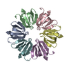 1u1tS S: Starting model for refinement C: citing same article ( |
|---|---|
| Similar structure data |
- Links
Links
- Assembly
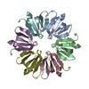
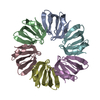
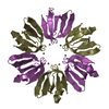
| Deposited unit | 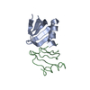
| ||||||||||||||||||
|---|---|---|---|---|---|---|---|---|---|---|---|---|---|---|---|---|---|---|---|
| 1 | 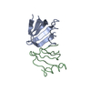
| ||||||||||||||||||
| Unit cell |
| ||||||||||||||||||
| Noncrystallographic symmetry (NCS) | NCS domain:
NCS domain segments: Component-ID: 1 / Ens-ID: 1 / Beg auth comp-ID: SER / Beg label comp-ID: SER / End auth comp-ID: LYS / End label comp-ID: LYS / Refine code: 2 / Auth seq-ID: 8 - 70 / Label seq-ID: 9 - 71
|
- Components
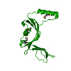
| #1: Protein | Mass: 8006.216 Da / Num. of mol.: 2 Source method: isolated from a genetically manipulated source Details: codons optimised for E. coli expression / Source: (gene. exp.)  Nostoc sp. (bacteria) / Strain: PCC 7120 / Gene: asl2047 / Plasmid: pTYP11 / Production host: Nostoc sp. (bacteria) / Strain: PCC 7120 / Gene: asl2047 / Plasmid: pTYP11 / Production host:  |
|---|
-Experimental details
-Experiment
| Experiment | Method:  X-RAY DIFFRACTION / Number of used crystals: 1 X-RAY DIFFRACTION / Number of used crystals: 1 |
|---|
- Sample preparation
| Crystal | Density Matthews: 2.15 Å3/Da / Density % sol: 42.74 % |
|---|---|
| Crystal grow | Temperature: 277 K / Method: vapor diffusion, sitting drop / pH: 3.5 Details: 2 M ammonium sulphate, 0.1 M citric acid, pH 3.5, VAPOR DIFFUSION, SITTING DROP, temperature 277K |
-Data collection
| Diffraction | Mean temperature: 100 K | ||||||||||||||||||||
|---|---|---|---|---|---|---|---|---|---|---|---|---|---|---|---|---|---|---|---|---|---|
| Diffraction source | Source:  SYNCHROTRON / Site: SYNCHROTRON / Site:  SLS SLS  / Beamline: X06SA / Wavelength: 0.9184 Å / Beamline: X06SA / Wavelength: 0.9184 Å | ||||||||||||||||||||
| Detector | Type: PSI PILATUS 6M / Detector: PIXEL / Date: Mar 3, 2008 | ||||||||||||||||||||
| Radiation | Monochromator: LN2 cooled fixed-exit Si(111) / Protocol: SINGLE WAVELENGTH / Monochromatic (M) / Laue (L): M / Scattering type: x-ray | ||||||||||||||||||||
| Radiation wavelength | Wavelength: 0.9184 Å / Relative weight: 1 | ||||||||||||||||||||
| Reflection twin |
| ||||||||||||||||||||
| Reflection | Resolution: 2.3→30.3 Å / Num. obs: 5806 / % possible obs: 97.9 % / Redundancy: 3.5 % / Biso Wilson estimate: 27.606 Å2 / Rmerge(I) obs: 0.065 / Net I/σ(I): 14.55 | ||||||||||||||||||||
| Reflection shell | Resolution: 2.3→2.4 Å / Redundancy: 3.4 % / Rmerge(I) obs: 0.179 / Mean I/σ(I) obs: 6.1 / Num. measured obs: 2232 / Num. unique obs: 648 / % possible all: 90.6 |
-Phasing
| Phasing | Method:  molecular replacement molecular replacement | |||||||||
|---|---|---|---|---|---|---|---|---|---|---|
| Phasing MR | Rfactor: 50.51
|
- Processing
| Software |
| |||||||||||||||||||||||||||||||||||||||||||||||||||||||||||||||||
|---|---|---|---|---|---|---|---|---|---|---|---|---|---|---|---|---|---|---|---|---|---|---|---|---|---|---|---|---|---|---|---|---|---|---|---|---|---|---|---|---|---|---|---|---|---|---|---|---|---|---|---|---|---|---|---|---|---|---|---|---|---|---|---|---|---|---|
| Refinement | Method to determine structure:  MOLECULAR REPLACEMENT MOLECULAR REPLACEMENTStarting model: PDB entry 1U1T Resolution: 2.31→30.29 Å / Cor.coef. Fo:Fc: 0.859 / Cor.coef. Fo:Fc free: 0.85 / WRfactor Rfree: 0.266 / WRfactor Rwork: 0.259 / Occupancy max: 1 / Occupancy min: 1 / FOM work R set: 0.667 / SU B: 11.773 / SU ML: 0.275 / SU R Cruickshank DPI: 0.099 / SU Rfree: 0.059 / Cross valid method: THROUGHOUT / σ(F): 0 / ESU R: 0.099 / ESU R Free: 0.059 / Stereochemistry target values: MAXIMUM LIKELIHOOD Details: HYDROGENS HAVE BEEN ADDED IN THE RIDING POSITIONS U VALUES : REFINED INDIVIDUALLY
| |||||||||||||||||||||||||||||||||||||||||||||||||||||||||||||||||
| Solvent computation | Ion probe radii: 0.8 Å / Shrinkage radii: 0.8 Å / VDW probe radii: 1.4 Å / Solvent model: BABINET MODEL WITH MASK | |||||||||||||||||||||||||||||||||||||||||||||||||||||||||||||||||
| Displacement parameters | Biso max: 42.01 Å2 / Biso mean: 18.63 Å2 / Biso min: 2 Å2
| |||||||||||||||||||||||||||||||||||||||||||||||||||||||||||||||||
| Refinement step | Cycle: LAST / Resolution: 2.31→30.29 Å
| |||||||||||||||||||||||||||||||||||||||||||||||||||||||||||||||||
| Refine LS restraints |
| |||||||||||||||||||||||||||||||||||||||||||||||||||||||||||||||||
| Refine LS restraints NCS | Dom-ID: 1 / Ens-ID: 1 / Refine-ID: X-RAY DIFFRACTION
| |||||||||||||||||||||||||||||||||||||||||||||||||||||||||||||||||
| LS refinement shell | Resolution: 2.305→2.365 Å / Total num. of bins used: 20
|
 Movie
Movie Controller








 PDBj
PDBj

