 ムービー
ムービー コントローラー


+ データを開く

- 基本情報
基本情報
| 登録情報 | データベース: PDB / ID: 3hfo | ||||||
|---|---|---|---|---|---|---|---|
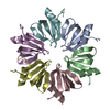
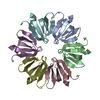
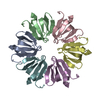
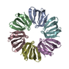
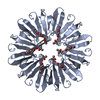
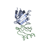
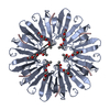
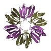
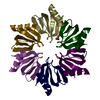
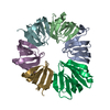
| タイトル | Crystal Structure of an Hfq protein from Synechocystis sp. | ||||||
 要素 要素 | Ssr3341 protein | ||||||
 キーワード キーワード | RNA BINDING PROTEIN / HFQ / SM / RNA-BINDING PROTEIN / SRNA / TRANSLATIONAL REGULATION | ||||||
| 機能・相同性 | : / Hfq related / SH3 type barrels. - #100 / LSM domain superfamily / SH3 type barrels. / Roll / Mainly Beta / Ssr3341 protein 機能・相同性情報 機能・相同性情報 | ||||||
| 生物種 |  | ||||||
| 手法 |  X線回折 / シンクロトロン / 分子置換 / 解像度: 1.3 Å X線回折 / シンクロトロン / 分子置換 / 解像度: 1.3 Å | ||||||
 データ登録者 データ登録者 | Boggild, A. / Overgaard, M. / Valentin-Hansen, P. / Brodersen, D.E. | ||||||
 引用 引用 | ジャーナル: Febs J. / 年: 2009 タイトル: Cyanobacteria contain a structural homologue of the Hfq protein with altered RNA-binding properties. 著者: Boggild, A. / Overgaard, M. / Valentin-Hansen, P. / Brodersen, D.E. | ||||||
| 履歴 |
|
- 構造の表示
構造の表示
| 構造ビューア | 分子: MolmilJmol/JSmol |
|---|
- ダウンロードとリンク
-ダウンロード
| PDBx/mmCIF形式 | 3hfo.cif.gz | 104.5 KB | 表示 | PDBx/mmCIF形式 |
|---|---|---|---|---|
| PDB形式 | pdb3hfo.ent.gz | 81.1 KB | 表示 | PDB形式 |
| PDBx/mmJSON形式 | 3hfo.json.gz | ツリー表示 | PDBx/mmJSON形式 | |
| その他 |  その他のダウンロード その他のダウンロード |
-検証レポート
| アーカイブディレクトリ | https://data.pdbj.org/pub/pdb/validation_reports/hf/3hfoftp://data.pdbj.org/pub/pdb/validation_reports/hf/3hfo | HTTPS FTP |
|---|
-関連構造データ


-リンク
 PDBj
PDBj

- 集合体
集合体
| 登録構造単位 | 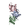
| ||||||||||||
|---|---|---|---|---|---|---|---|---|---|---|---|---|---|
| 1 | 
| ||||||||||||
| 単位格子 |
| ||||||||||||
| Components on special symmetry positions |
|
-要素
| #1: タンパク質 | 分子量: 7839.961 Da / 分子数: 3 / 由来タイプ: 組換発現 / 詳細: codons optimised for E. coli expression 由来: (組換発現) 株: PCC6803 / 遺伝子: ssr3341 / プラスミド: pTYP11 / 発現宿主: #2: 水 | ChemComp-HOH / |  分子量: 18.015 Da / 分子数: 272 / 由来タイプ: 天然 / 式: H2O 分子量: 18.015 Da / 分子数: 272 / 由来タイプ: 天然 / 式: H2O |
|---|
-実験情報
-実験
| 実験 | 手法: X線回折 / 使用した結晶の数: 1 |
|---|
- 試料調製
試料調製
| 結晶 | マシュー密度: 1.92 Å3/Da / 溶媒含有率: 36.09 % |
|---|---|
| 結晶化 | 温度: 292 K / 手法: 蒸気拡散法, シッティングドロップ法 / pH: 7 詳細: 60% Tacsimate, pH 7.0, VAPOR DIFFUSION, SITTING DROP, temperature 292K |
-データ収集
| 回折 | 平均測定温度: 100 K | |||||||||||||||||||||||||||||||||||||||||||||||||||||||||||||||||||||||||||||||||||||||||||||||||||||||||||||||||||||||||||||||||||||||||||||||||||
|---|---|---|---|---|---|---|---|---|---|---|---|---|---|---|---|---|---|---|---|---|---|---|---|---|---|---|---|---|---|---|---|---|---|---|---|---|---|---|---|---|---|---|---|---|---|---|---|---|---|---|---|---|---|---|---|---|---|---|---|---|---|---|---|---|---|---|---|---|---|---|---|---|---|---|---|---|---|---|---|---|---|---|---|---|---|---|---|---|---|---|---|---|---|---|---|---|---|---|---|---|---|---|---|---|---|---|---|---|---|---|---|---|---|---|---|---|---|---|---|---|---|---|---|---|---|---|---|---|---|---|---|---|---|---|---|---|---|---|---|---|---|---|---|---|---|---|---|---|
| 放射光源 | 由来: シンクロトロン / サイト: MAX II  / ビームライン: I911-3 / 波長: 0.9 Å / ビームライン: I911-3 / 波長: 0.9 Å | |||||||||||||||||||||||||||||||||||||||||||||||||||||||||||||||||||||||||||||||||||||||||||||||||||||||||||||||||||||||||||||||||||||||||||||||||||
| 検出器 | タイプ: MARMOSAIC 225 mm CCD / 検出器: CCD / 日付: 2008年6月19日 | |||||||||||||||||||||||||||||||||||||||||||||||||||||||||||||||||||||||||||||||||||||||||||||||||||||||||||||||||||||||||||||||||||||||||||||||||||
| 放射 | モノクロメーター: Double crystal monochromator, Si(111) プロトコル: SINGLE WAVELENGTH / 単色(M)・ラウエ(L): M / 散乱光タイプ: x-ray | |||||||||||||||||||||||||||||||||||||||||||||||||||||||||||||||||||||||||||||||||||||||||||||||||||||||||||||||||||||||||||||||||||||||||||||||||||
| 放射波長 | 波長: 0.9 Å / 相対比: 1 | |||||||||||||||||||||||||||||||||||||||||||||||||||||||||||||||||||||||||||||||||||||||||||||||||||||||||||||||||||||||||||||||||||||||||||||||||||
| 反射 | 解像度: 1.21→26.2 Å / Num. obs: 52709 / % possible obs: 96.5 % / 冗長度: 4.8 % / Biso Wilson estimate: 16.864 Å2 / Rmerge(I) obs: 0.048 / Net I/σ(I): 24.23 | |||||||||||||||||||||||||||||||||||||||||||||||||||||||||||||||||||||||||||||||||||||||||||||||||||||||||||||||||||||||||||||||||||||||||||||||||||
| 反射 シェル | Diffraction-ID: 1
|
-位相決定
| 位相決定 | 手法: 分子置換 | |||||||||
|---|---|---|---|---|---|---|---|---|---|---|
| Phasing MR | Rfactor: 51.73
|
- 解析
解析
| ソフトウェア |
| |||||||||||||||||||||||||||||||||||||||||||||||||||||||||||||||||||||||||||||||||||||||||||
|---|---|---|---|---|---|---|---|---|---|---|---|---|---|---|---|---|---|---|---|---|---|---|---|---|---|---|---|---|---|---|---|---|---|---|---|---|---|---|---|---|---|---|---|---|---|---|---|---|---|---|---|---|---|---|---|---|---|---|---|---|---|---|---|---|---|---|---|---|---|---|---|---|---|---|---|---|---|---|---|---|---|---|---|---|---|---|---|---|---|---|---|---|
| 精密化 | 構造決定の手法: 分子置換 開始モデル: PDB entry 1HK9 解像度: 1.3→26.199 Å / Occupancy max: 1 / Occupancy min: 0.16 / FOM work R set: 0.891 / SU ML: 0.04 / 立体化学のターゲット値: ML
| |||||||||||||||||||||||||||||||||||||||||||||||||||||||||||||||||||||||||||||||||||||||||||
| 溶媒の処理 | 減衰半径: 0.9 Å / VDWプローブ半径: 1.11 Å / 溶媒モデル: FLAT BULK SOLVENT MODEL / Bsol: 56.911 Å2 / ksol: 0.389 e/Å3 | |||||||||||||||||||||||||||||||||||||||||||||||||||||||||||||||||||||||||||||||||||||||||||
| 原子変位パラメータ | Biso max: 64.74 Å2 / Biso mean: 17.734 Å2 / Biso min: 5.63 Å2
| |||||||||||||||||||||||||||||||||||||||||||||||||||||||||||||||||||||||||||||||||||||||||||
| 精密化ステップ | サイクル: LAST / 解像度: 1.3→26.199 Å
| |||||||||||||||||||||||||||||||||||||||||||||||||||||||||||||||||||||||||||||||||||||||||||
| 拘束条件 |
| |||||||||||||||||||||||||||||||||||||||||||||||||||||||||||||||||||||||||||||||||||||||||||
| LS精密化 シェル | Refine-ID: X-RAY DIFFRACTION / Total num. of bins used: 12
|
