 Movie
Movie Controller


+ Open data

- Basic information
| Entry | Database: PDB / ID: 1kq2 | ||||||
|---|---|---|---|---|---|---|---|
| Title | Crystal Structure of an Hfq-RNA Complex | ||||||
 Components Components |
| ||||||
 Keywords Keywords | TRANSLATION/RNA / Hfq-RNA complex / single-stranded RNA / translational regulator / TRANSLATION-RNA COMPLEX | ||||||
| Function / homology |  Function and homology information Function and homology informationregulation of translation, ncRNA-mediated / regulation of RNA stability / regulation of DNA-templated transcription / RNA binding / cytosol Similarity search - Function | ||||||
| Biological species |   Staphylococcus aureus (bacteria) Staphylococcus aureus (bacteria) | ||||||
| Method |  X-RAY DIFFRACTION / SYNCHROTRON / MOLECULAR REPLACEMENT / Resolution: 2.71 Å X-RAY DIFFRACTION / SYNCHROTRON / MOLECULAR REPLACEMENT / Resolution: 2.71 Å | ||||||
 Authors Authors | Schumacher, M.A. / Pearson, R.F. / Moller, T. / Valentin-Hansen, P. / Brennan, R.G. | ||||||
 Citation Citation | Journal: EMBO J. / Year: 2002 Title: Structures of the pleiotropic translational regulator Hfq and an Hfq-RNA complex: a bacterial Sm-like protein. Authors: Schumacher, M.A. / Pearson, R.F. / Moller, T. / Valentin-Hansen, P. / Brennan, R.G. | ||||||
| History |
|
- Structure visualization
| Structure viewer | Molecule: MolmilJmol/JSmol |
|---|
- Downloads & links
-Download
| PDBx/mmCIF format | 1kq2.cif.gz | 88.2 KB | Display | PDBx/mmCIF format |
|---|---|---|---|---|
| PDB format | pdb1kq2.ent.gz | 67.9 KB | Display | PDB format |
| PDBx/mmJSON format | 1kq2.json.gz | Tree view | PDBx/mmJSON format | |
| Others |  Other downloads Other downloads |
-Validation report
| Arichive directory | https://data.pdbj.org/pub/pdb/validation_reports/kq/1kq2ftp://data.pdbj.org/pub/pdb/validation_reports/kq/1kq2 | HTTPS FTP |
|---|
-Related structure data
| Related structure data |  1kq1SC S: Starting model for refinement C: citing same article ( |
|---|---|
| Similar structure data |


-Links
 PDBj
PDBj

- Assembly
| Deposited unit | 
| ||||||||
|---|---|---|---|---|---|---|---|---|---|
| 1 |
| ||||||||
| Unit cell |
| ||||||||
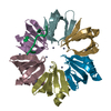
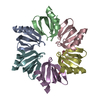
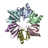
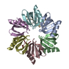
| Details | Hfq is a functional hexamer and there is one hexamer bound to the 7-mer RNA site in the ASU |
-Components
| #1: RNA chain | Mass: 2160.283 Da / Num. of mol.: 1 / Source method: obtained synthetically | ||
|---|---|---|---|
| #2: Protein | Mass: 8786.683 Da / Num. of mol.: 6 Source method: isolated from a genetically manipulated source Source: (gene. exp.) Staphylococcus aureus (bacteria) / Plasmid: pTyb11 / Production host: #3: Water | ChemComp-HOH / |  Mass: 18.015 Da / Num. of mol.: 29 / Source method: isolated from a natural source / Formula: H2O Mass: 18.015 Da / Num. of mol.: 29 / Source method: isolated from a natural source / Formula: H2O |
-Experimental details
-Experiment
| Experiment | Method: X-RAY DIFFRACTION / Number of used crystals: 1 |
|---|
- Sample preparation
| Crystal | Density Matthews: 2.17 Å3/Da / Density % sol: 43.28 % | |||||||||||||||||||||||||||||||||||||||||||||||||
|---|---|---|---|---|---|---|---|---|---|---|---|---|---|---|---|---|---|---|---|---|---|---|---|---|---|---|---|---|---|---|---|---|---|---|---|---|---|---|---|---|---|---|---|---|---|---|---|---|---|---|
| Crystal grow | Temperature: 298 K / Method: vapor diffusion, hanging drop / pH: 7.5 Details: PEG 550, MgCl2, Hepes, KCl, pH 7.5, VAPOR DIFFUSION, HANGING DROP, temperature 298K | |||||||||||||||||||||||||||||||||||||||||||||||||
| Components of the solutions |
| |||||||||||||||||||||||||||||||||||||||||||||||||
| Crystal grow | *PLUS Method: microdialysis | |||||||||||||||||||||||||||||||||||||||||||||||||
| Components of the solutions | *PLUS
|
-Data collection
| Diffraction | Mean temperature: 298 K |
|---|---|
| Diffraction source | Source: SYNCHROTRON / Site: SSRL  / Beamline: BL9-1 / Wavelength: 0.97 / Beamline: BL9-1 / Wavelength: 0.97 |
| Detector | Type: MARRESEARCH / Detector: IMAGE PLATE / Date: Jun 11, 2001 |
| Radiation | Monochromator: graphite / Protocol: SINGLE WAVELENGTH / Monochromatic (M) / Laue (L): M / Scattering type: x-ray |
| Radiation wavelength | Wavelength: 0.97 Å / Relative weight: 1 |
| Reflection | Resolution: 2.71→40.38 Å / Num. all: 11950 / Num. obs: 11950 / % possible obs: 90.3 % / Observed criterion σ(F): 0 / Observed criterion σ(I): 0 / Biso Wilson estimate: 83.4 Å2 / Rsym value: 0.064 |
| Reflection shell | Resolution: 2.71→2.89 Å / % possible all: 92.1 |
| Reflection | *PLUS Num. measured all: 40717 / Rmerge(I) obs: 0.064 |
| Reflection shell | *PLUS Lowest resolution: 2.8 Å / Rmerge(I) obs: 0.367 / Mean I/σ(I) obs: 2 |
- Processing
| Software |
| ||||||||||||||||||||||||||||||||||||
|---|---|---|---|---|---|---|---|---|---|---|---|---|---|---|---|---|---|---|---|---|---|---|---|---|---|---|---|---|---|---|---|---|---|---|---|---|---|
| Refinement | Method to determine structure: MOLECULAR REPLACEMENT Starting model: one hexamer of 1KQ1 Resolution: 2.71→40.38 Å / Rfactor Rfree error: 0.011 / Data cutoff high absF: 2081750.71 / Data cutoff low absF: 0 / Isotropic thermal model: RESTRAINED / Cross valid method: THROUGHOUT / σ(F): 0 / Stereochemistry target values: engh and huber
| ||||||||||||||||||||||||||||||||||||
| Solvent computation | Solvent model: FLAT MODEL / Bsol: 52.977 Å2 / ksol: 0.323089 e/Å3 | ||||||||||||||||||||||||||||||||||||
| Displacement parameters | Biso mean: 64.5 Å2
| ||||||||||||||||||||||||||||||||||||
| Refine analyze |
| ||||||||||||||||||||||||||||||||||||
| Refinement step | Cycle: LAST / Resolution: 2.71→40.38 Å
| ||||||||||||||||||||||||||||||||||||
| Refine LS restraints |
| ||||||||||||||||||||||||||||||||||||
| LS refinement shell | Resolution: 2.71→2.88 Å / Rfactor Rfree error: 0.037 / Total num. of bins used: 6
| ||||||||||||||||||||||||||||||||||||
| Xplor file |
| ||||||||||||||||||||||||||||||||||||
| Refinement | *PLUS Lowest resolution: 40.4 Å / % reflection Rfree: 5 % / Rfactor obs: 0.204 / Rfactor Rfree: 0.266 / Rfactor Rwork: 0.204 | ||||||||||||||||||||||||||||||||||||
| Solvent computation | *PLUS | ||||||||||||||||||||||||||||||||||||
| Displacement parameters | *PLUS | ||||||||||||||||||||||||||||||||||||
| Refine LS restraints | *PLUS
| ||||||||||||||||||||||||||||||||||||
| LS refinement shell | *PLUS Rfactor Rfree: 0.377 / Rfactor Rwork: 0.315 |
