 Movie
Movie Controller


+ Open data

- Basic information
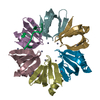
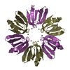
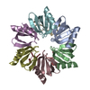
| Entry | Database: PDB / ID: 4qvc | ||||||
|---|---|---|---|---|---|---|---|
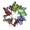
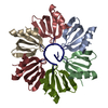
| Title | E.coli Hfq in complex with RNA Aus | ||||||
 Components Components |
| ||||||
 Keywords Keywords | RNA binding protein/RNA / SM fold / RNA binding / RNA / RNA binding protein-RNA complex | ||||||
| Function / homology |  Function and homology information Function and homology informationregulation of translation, ncRNA-mediated / regulation of RNA stability / regulation of DNA-templated transcription / RNA binding / cytosol Similarity search - Function | ||||||
| Biological species |  | ||||||
| Method |  X-RAY DIFFRACTION / SYNCHROTRON / MOLECULAR REPLACEMENT / Resolution: 1.99 Å X-RAY DIFFRACTION / SYNCHROTRON / MOLECULAR REPLACEMENT / Resolution: 1.99 Å | ||||||
 Authors Authors | Wang, L.J. / Wang, W.W. / Li, F.D. / Wu, J.H. / Gong, Q.G. / Shi, Y.Y. | ||||||
 Citation Citation | Journal: Nucleic Acids Res. / Year: 2015 Title: Structural insights into the recognition of the internal A-rich linker from OxyS sRNA by Escherichia coli Hfq Authors: Wang, L.J. / Wang, W.W. / Li, F.D. / Zhang, J. / Wu, J.H. / Gong, Q.G. / Shi, Y.Y. | ||||||
| History |
|
- Structure visualization
| Structure viewer | Molecule: MolmilJmol/JSmol |
|---|
- Downloads & links
-Download
| PDBx/mmCIF format | 4qvc.cif.gz | 89.8 KB | Display | PDBx/mmCIF format |
|---|---|---|---|---|
| PDB format | pdb4qvc.ent.gz | 67.1 KB | Display | PDB format |
| PDBx/mmJSON format | 4qvc.json.gz | Tree view | PDBx/mmJSON format | |
| Others |  Other downloads Other downloads |
-Validation report
| Arichive directory | https://data.pdbj.org/pub/pdb/validation_reports/qv/4qvcftp://data.pdbj.org/pub/pdb/validation_reports/qv/4qvc | HTTPS FTP |
|---|
-Related structure data
| Related structure data | 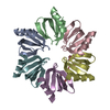 4qvdC 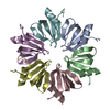 1hk9S C: citing same article ( S: Starting model for refinement |
|---|---|
| Similar structure data |


-Links
 PDBj
PDBj

- Assembly
| Deposited unit | 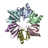
| ||||||||
|---|---|---|---|---|---|---|---|---|---|
| 1 |
| ||||||||
| Unit cell |
|
-Components
| #1: Protein | Mass: 7325.554 Da / Num. of mol.: 6 / Fragment: UNP residues 1-65 Source method: isolated from a genetically manipulated source Source: (gene. exp.) #2: RNA chain | | Mass: 2189.379 Da / Num. of mol.: 1 / Source method: obtained synthetically / Details: This sequence occurs naturally in E.coli. / Source: (synth.) #3: Water | ChemComp-HOH / |  Mass: 18.015 Da / Num. of mol.: 183 / Source method: isolated from a natural source / Formula: H2O Mass: 18.015 Da / Num. of mol.: 183 / Source method: isolated from a natural source / Formula: H2O |
|---|
-Experimental details
-Experiment
| Experiment | Method: X-RAY DIFFRACTION / Number of used crystals: 1 |
|---|
- Sample preparation
| Crystal | Density Matthews: 2.43 Å3/Da / Density % sol: 49.3 % / Mosaicity: 0.554 ° |
|---|---|
| Crystal grow | Temperature: 281 K / Method: vapor diffusion, hanging drop / pH: 5.5 Details: 12% PEG4000, 0.1M citrate, pH 5.5, VAPOR DIFFUSION, HANGING DROP, temperature 281K |
-Data collection
| Diffraction | Mean temperature: 100 K | ||||||||||||||||||||||||||||||||||||||||||||||||||||||||||||||||||
|---|---|---|---|---|---|---|---|---|---|---|---|---|---|---|---|---|---|---|---|---|---|---|---|---|---|---|---|---|---|---|---|---|---|---|---|---|---|---|---|---|---|---|---|---|---|---|---|---|---|---|---|---|---|---|---|---|---|---|---|---|---|---|---|---|---|---|---|
| Diffraction source | Source: SYNCHROTRON / Site: SSRF  / Beamline: BL17U / Wavelength: 0.97923 Å / Beamline: BL17U / Wavelength: 0.97923 Å | ||||||||||||||||||||||||||||||||||||||||||||||||||||||||||||||||||
| Detector | Type: ADSC QUANTUM 315r / Detector: CCD / Date: Apr 10, 2013 | ||||||||||||||||||||||||||||||||||||||||||||||||||||||||||||||||||
| Radiation | Monochromator: Si 111 double crystal / Protocol: SINGLE WAVELENGTH / Monochromatic (M) / Laue (L): M / Scattering type: x-ray | ||||||||||||||||||||||||||||||||||||||||||||||||||||||||||||||||||
| Radiation wavelength | Wavelength: 0.97923 Å / Relative weight: 1 | ||||||||||||||||||||||||||||||||||||||||||||||||||||||||||||||||||
| Reflection | Resolution: 1.99→40 Å / Num. all: 31605 / Num. obs: 29582 / % possible obs: 93.6 % / Observed criterion σ(F): 1 / Observed criterion σ(I): 1 / Redundancy: 8.1 % / Rmerge(I) obs: 0.105 / Χ2: 1.331 / Net I/σ(I): 11.1 | ||||||||||||||||||||||||||||||||||||||||||||||||||||||||||||||||||
| Reflection shell | Diffraction-ID: 1 / Rejects: _
|
- Processing
| Software |
| |||||||||||||||||||||||||||||||||||||||||||||||||||||||||||||||||||||||||||
|---|---|---|---|---|---|---|---|---|---|---|---|---|---|---|---|---|---|---|---|---|---|---|---|---|---|---|---|---|---|---|---|---|---|---|---|---|---|---|---|---|---|---|---|---|---|---|---|---|---|---|---|---|---|---|---|---|---|---|---|---|---|---|---|---|---|---|---|---|---|---|---|---|---|---|---|---|
| Refinement | Method to determine structure: MOLECULAR REPLACEMENT Starting model: 1HK9 Resolution: 1.99→33.99 Å / Cor.coef. Fo:Fc: 0.948 / Cor.coef. Fo:Fc free: 0.937 / WRfactor Rfree: 0.2629 / WRfactor Rwork: 0.2141 / FOM work R set: 0.8037 / SU B: 4.6 / SU ML: 0.126 / SU R Cruickshank DPI: 0.1964 / SU Rfree: 0.1745 / Cross valid method: THROUGHOUT / σ(F): 0 / ESU R: 0.196 / ESU R Free: 0.175 / Stereochemistry target values: MAXIMUM LIKELIHOOD Details: HYDROGENS HAVE BEEN ADDED IN THE RIDING POSITIONS U VALUES: REFINED INDIVIDUALLY
| |||||||||||||||||||||||||||||||||||||||||||||||||||||||||||||||||||||||||||
| Solvent computation | Ion probe radii: 0.8 Å / Shrinkage radii: 0.8 Å / VDW probe radii: 1.2 Å / Solvent model: MASK | |||||||||||||||||||||||||||||||||||||||||||||||||||||||||||||||||||||||||||
| Displacement parameters | Biso max: 81.64 Å2 / Biso mean: 34.715 Å2 / Biso min: 16.53 Å2
| |||||||||||||||||||||||||||||||||||||||||||||||||||||||||||||||||||||||||||
| Refinement step | Cycle: LAST / Resolution: 1.99→33.99 Å
| |||||||||||||||||||||||||||||||||||||||||||||||||||||||||||||||||||||||||||
| Refine LS restraints |
| |||||||||||||||||||||||||||||||||||||||||||||||||||||||||||||||||||||||||||
| LS refinement shell | Resolution: 1.988→2.04 Å / Total num. of bins used: 20
|
