 Movie
Movie Controller

[English] 日本語
 Yorodumi
- PDB-1kq1: 1.55 A Crystal structure of the pleiotropic translational regulat... -
+ Open data

- Basic information
| Entry | Database: PDB / ID: 1kq1 | ||||||
|---|---|---|---|---|---|---|---|
| Title | 1.55 A Crystal structure of the pleiotropic translational regulator, Hfq | ||||||
 Components Components | Host Factor for Q beta | ||||||
 Keywords Keywords | TRANSLATION / Hfq / hexamer / RNA binding protein / translational regulator / Sm motif | ||||||
| Function / homology |  Function and homology information Function and homology informationregulation of translation, ncRNA-mediated / regulation of RNA stability / regulation of DNA-templated transcription / RNA binding / cytosol Similarity search - Function | ||||||
| Biological species |   Staphylococcus aureus (bacteria) Staphylococcus aureus (bacteria) | ||||||
| Method |  X-RAY DIFFRACTION / SYNCHROTRON / SIR / Resolution: 1.55 Å X-RAY DIFFRACTION / SYNCHROTRON / SIR / Resolution: 1.55 Å | ||||||
 Authors Authors | Schumacher, M.A. / Pearson, R.F. / Moller, T. / Valentin-Hansen, P. / Brennan, R.G. | ||||||
 Citation Citation | Journal: EMBO J. / Year: 2002 Title: Structures of the pleiotropic translational regulator Hfq and an Hfq-RNA complex: a bacterial Sm-like protein. Authors: Schumacher, M.A. / Pearson, R.F. / Moller, T. / Valentin-Hansen, P. / Brennan, R.G. | ||||||
| History |
|
- Structure visualization
| Structure viewer | Molecule: MolmilJmol/JSmol |
|---|
- Downloads & links
-Download
| PDBx/mmCIF format | 1kq1.cif.gz | 167.9 KB | Display | PDBx/mmCIF format |
|---|---|---|---|---|
| PDB format | pdb1kq1.ent.gz | 136.4 KB | Display | PDB format |
| PDBx/mmJSON format | 1kq1.json.gz | Tree view | PDBx/mmJSON format | |
| Others |  Other downloads Other downloads |
-Validation report
| Arichive directory | https://data.pdbj.org/pub/pdb/validation_reports/kq/1kq1ftp://data.pdbj.org/pub/pdb/validation_reports/kq/1kq1 | HTTPS FTP |
|---|
-Related structure data


-Links
 PDBj
PDBj


- Assembly
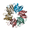
| Deposited unit | 
| ||||||||
|---|---|---|---|---|---|---|---|---|---|
| 1 | 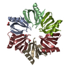
| ||||||||
| 2 | 
| ||||||||
| Unit cell |
| ||||||||
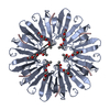
| Details | Hfq forms a hexamer (resistant to denaturants) and there are two hexamers in the crystallographic asymmetric unit |
-Components
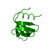
| #1: Protein | Mass: 8786.683 Da / Num. of mol.: 12 Source method: isolated from a genetically manipulated source Source: (gene. exp.) Staphylococcus aureus (bacteria) / Production host: #2: Chemical | ChemComp-ACY /   Mass: 60.052 Da / Num. of mol.: 5 / Source method: obtained synthetically / Formula: C2H4O2 Mass: 60.052 Da / Num. of mol.: 5 / Source method: obtained synthetically / Formula: C2H4O2#3: Water | ChemComp-HOH / |  Mass: 18.015 Da / Num. of mol.: 592 / Source method: isolated from a natural source / Formula: H2O Mass: 18.015 Da / Num. of mol.: 592 / Source method: isolated from a natural source / Formula: H2O |
|---|
-Experimental details
-Experiment
| Experiment | Method: X-RAY DIFFRACTION / Number of used crystals: 1 |
|---|
- Sample preparation
| Crystal | Density Matthews: 1.92 Å3/Da / Density % sol: 35.9 % | |||||||||||||||||||||||||||||||||||||||||||||||||
|---|---|---|---|---|---|---|---|---|---|---|---|---|---|---|---|---|---|---|---|---|---|---|---|---|---|---|---|---|---|---|---|---|---|---|---|---|---|---|---|---|---|---|---|---|---|---|---|---|---|---|
| Crystal grow | Temperature: 298 K / Method: vapor diffusion, hanging drop / pH: 4.6 Details: ammonium sulphate, sodium acetate, pH 4.6, VAPOR DIFFUSION, HANGING DROP, temperature 298K | |||||||||||||||||||||||||||||||||||||||||||||||||
| Crystal grow | *PLUS pH: 7.5 / Method: microdialysis | |||||||||||||||||||||||||||||||||||||||||||||||||
| Components of the solutions | *PLUS
|
-Data collection
| Diffraction | Mean temperature: 100 K |
|---|---|
| Diffraction source | Source: SYNCHROTRON / Site: SSRL  / Beamline: BL9-1 / Wavelength: 0.97 Å / Beamline: BL9-1 / Wavelength: 0.97 Å |
| Detector | Type: MARRESEARCH / Detector: IMAGE PLATE / Date: May 11, 2001 |
| Radiation | Monochromator: graphite / Protocol: SINGLE WAVELENGTH / Monochromatic (M) / Laue (L): M / Scattering type: x-ray |
| Radiation wavelength | Wavelength: 0.97 Å / Relative weight: 1 |
| Reflection | Resolution: 1.55→22.37 Å / Num. obs: 115018 / % possible obs: 99.8 % / Observed criterion σ(F): 0 / Observed criterion σ(I): 0 / Biso Wilson estimate: 22.7 Å2 / Rsym value: 0.046 / Net I/σ(I): 7.6 |
| Reflection shell | Resolution: 1.55→1.65 Å / Rsym value: 0.158 / % possible all: 100 |
| Reflection | *PLUS Lowest resolution: 22.4 Å / Num. obs: 111857 / Num. measured all: 460147 / Rmerge(I) obs: 0.046 |
| Reflection shell | *PLUS Lowest resolution: 1.6 Å / Rmerge(I) obs: 0.158 / Mean I/σ(I) obs: 4.7 |
- Processing
| Software |
| ||||||||||||||||||||||||
|---|---|---|---|---|---|---|---|---|---|---|---|---|---|---|---|---|---|---|---|---|---|---|---|---|---|
| Refinement | Method to determine structure: SIR / Resolution: 1.55→22.37 Å / Rfactor Rfree error: 0.003 / Data cutoff high absF: 1029855.35 / Data cutoff low absF: 0 / Isotropic thermal model: RESTRAINED / Cross valid method: THROUGHOUT / σ(F): 0 / Stereochemistry target values: Engh and Huber Details: residues 67 to 77 are disordered in all subunits as are residues 1-5 in 11 subunits.
| ||||||||||||||||||||||||
| Solvent computation | Solvent model: FLAT MODEL / Bsol: 63.3442 Å2 / ksol: 0.46278 e/Å3 | ||||||||||||||||||||||||
| Displacement parameters | Biso mean: 23.9 Å2
| ||||||||||||||||||||||||
| Refine analyze |
| ||||||||||||||||||||||||
| Refinement step | Cycle: LAST / Resolution: 1.55→22.37 Å
| ||||||||||||||||||||||||
| Refine LS restraints |
| ||||||||||||||||||||||||
| LS refinement shell | Resolution: 1.55→1.65 Å / Rfactor Rfree error: 0.012 / Total num. of bins used: 6
| ||||||||||||||||||||||||
| Xplor file |
| ||||||||||||||||||||||||
| Refinement | *PLUS Lowest resolution: 22.4 Å / % reflection Rfree: 5 % / Rfactor Rfree: 0.259 / Rfactor Rwork: 0.237 | ||||||||||||||||||||||||
| Solvent computation | *PLUS | ||||||||||||||||||||||||
| Displacement parameters | *PLUS | ||||||||||||||||||||||||
| Refine LS restraints | *PLUS
| ||||||||||||||||||||||||
| LS refinement shell | *PLUS Rfactor Rfree: 0.374 / Rfactor Rwork: 0.34 |
