 Movie
Movie Controller


+ Open data

- Basic information
| Entry | Database: PDB / ID: 1hk9 | ||||||
|---|---|---|---|---|---|---|---|
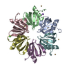
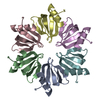
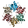
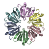
| Title | Crystal structure of the Hfq protein from Escherichia coli | ||||||
 Components Components | PROTEIN HFQ | ||||||
 Keywords Keywords | RNA BINDING PROTEIN / RNA-BINDING PROTEIN / SM-LIKE / PLEIOTROPIC REGULATOR / RNA CHAPERONE | ||||||
| Function / homology |  Function and homology information Function and homology informationsRNA-mediated post-transcriptional gene silencing / positive regulation of translation, ncRNA-mediated / bacterial nucleoid / regulation of translation, ncRNA-mediated / bent DNA binding / regulation of RNA stability / RNA folding chaperone / tRNA processing / tRNA binding / regulation of DNA-templated transcription ...sRNA-mediated post-transcriptional gene silencing / positive regulation of translation, ncRNA-mediated / bacterial nucleoid / regulation of translation, ncRNA-mediated / bent DNA binding / regulation of RNA stability / RNA folding chaperone / tRNA processing / tRNA binding / regulation of DNA-templated transcription / DNA binding / RNA binding / ATP binding / cytosol Similarity search - Function | ||||||
| Biological species |  | ||||||
| Method |  X-RAY DIFFRACTION / SYNCHROTRON / MOLECULAR REPLACEMENT / Resolution: 2.15 Å X-RAY DIFFRACTION / SYNCHROTRON / MOLECULAR REPLACEMENT / Resolution: 2.15 Å | ||||||
 Authors Authors | Sauter, C. / Basquin, J. / Suck, D. | ||||||
 Citation Citation | Journal: Nucleic Acids Res. / Year: 2003 Title: Sm-Like Proteins in Eubacteria: The Crystal Structure of the Hfq Protein from Escherichia Coli Authors: Sauter, C. / Basquin, J. / Suck, D. #1: Journal: Mol.Cell / Year: 2002 Title: The Sm-Like Hfq Protein Increases Oxys RNA Interaction with Target Mrnas Authors: Zhang, A. / Wassarman, K.M. / Ortega, J. / Steven, A.C. / Storz, G. #2: Journal: Mol.Cell / Year: 2002 Title: Hfq: A Bacterial Sm-Like Protein that Mediates RNA-RNA Interaction Authors: Moller, T. / Franch, T. / Hojrup, P. / Keene, D.R. / Bachinger, H.P. / Brennan, R.G. / Valentin-Hansen, P. #3: Journal: Embo J. / Year: 2002Title: Structures of the Pleiotropic Translational Regulator Hfq and an Hfq-RNA Complex: A Bacterial Sm-Like Protein Authors: Schumacher, M.A. / Pearson, R.F. / Moller, T. / Valentin-Hansen, P. / Brennan, R.G. #4: Journal: Mol.Microbiol. / Year: 1994 Title: Characterization of Broadly Pleiotropic Phenotypes Caused by an Hfq Insertion Mutation in Escherichia Coli K-12 Authors: Tsui, H.C. / Leung, H.C. / Winkler, M.E. #5: Journal: J.Biol.Chem. / Year: 1972 Title: Bacterial Proteins Required for Replication of Phage Q Ribonucleic Acid. Purification and Properties of Host Factor I, a Ribonucleic Acid-Binding Protein Authors: De Fernandez, M.T.F. / Hayward, W.S. / August, J.T. | ||||||
| History |
| ||||||
| Remark 650 | HELIX DETERMINATION METHOD: AUTHOR PROVIDED. | ||||||
| Remark 700 | SHEET DETERMINATION METHOD: AUTHOR PROVIDED. |
- Structure visualization
| Structure viewer | Molecule: MolmilJmol/JSmol |
|---|
- Downloads & links
-Download
| PDBx/mmCIF format | 1hk9.cif.gz | 90.6 KB | Display | PDBx/mmCIF format |
|---|---|---|---|---|
| PDB format | pdb1hk9.ent.gz | 69.9 KB | Display | PDB format |
| PDBx/mmJSON format | 1hk9.json.gz | Tree view | PDBx/mmJSON format | |
| Others |  Other downloads Other downloads |
-Validation report
| Arichive directory | https://data.pdbj.org/pub/pdb/validation_reports/hk/1hk9ftp://data.pdbj.org/pub/pdb/validation_reports/hk/1hk9 | HTTPS FTP |
|---|
-Related structure data
| Related structure data |  1kq1S S: Starting model for refinement |
|---|---|
| Similar structure data |


-Links
 PDBj
PDBj

- Assembly
| Deposited unit | 
| ||||||||||||||||||||||||
|---|---|---|---|---|---|---|---|---|---|---|---|---|---|---|---|---|---|---|---|---|---|---|---|---|---|
| 1 |
| ||||||||||||||||||||||||
| Unit cell |
| ||||||||||||||||||||||||
| Noncrystallographic symmetry (NCS) | NCS oper:
|
-Components
| #1: Protein | Mass: 8257.572 Da / Num. of mol.: 6 / Fragment: RESIDUES 1-72 Source method: isolated from a genetically manipulated source Details: C-TERMINAL RESIDUES 73-102 DELETED / Source: (gene. exp.) #2: Water | ChemComp-HOH / |  Mass: 18.015 Da / Num. of mol.: 136 / Source method: isolated from a natural source / Formula: H2O Mass: 18.015 Da / Num. of mol.: 136 / Source method: isolated from a natural source / Formula: H2OCompound details | RNA-BINDING PROTEIN THAT STIMULATES THE ELONGATION OF POLY(A) TAILS.EXISTS AS A HOMOHEXAMER. MAY ...RNA-BINDING PROTEIN THAT STIMULATES | |
|---|
-Experimental details
-Experiment
| Experiment | Method: X-RAY DIFFRACTION / Number of used crystals: 1 |
|---|
- Sample preparation
| Crystal | Density Matthews: 1.83 Å3/Da / Density % sol: 33 % | ||||||||||||||||||||||||||||||
|---|---|---|---|---|---|---|---|---|---|---|---|---|---|---|---|---|---|---|---|---|---|---|---|---|---|---|---|---|---|---|---|
| Crystal grow | Temperature: 293 K / Method: vapor diffusion, sitting drop / pH: 4.6 Details: CRYSTALS WERE OBTAINED BY VAPOR DIFFUSION IN 2UL SITTING DROPS. THE RESERVOIR CONTAINED 25% PEG 4000, 0.2 M NH4-ACETATE AND 0.2 M NA-ACETATE PH 4.6. CRYSTALLIZATION WERE CARRIED OUT AT 20C. | ||||||||||||||||||||||||||||||
| Crystal grow | *PLUS Temperature: 20 ℃ / pH: 4.6 / Method: vapor diffusion, hanging drop | ||||||||||||||||||||||||||||||
| Components of the solutions | *PLUS
|
-Data collection
| Diffraction | Mean temperature: 110 K |
|---|---|
| Diffraction source | Source: SYNCHROTRON / Site: ELETTRA  / Beamline: 5.2R / Wavelength: 1 / Beamline: 5.2R / Wavelength: 1 |
| Detector | Type: MARRESEARCH / Detector: CCD / Date: Jul 15, 2002 |
| Radiation | Monochromator: SILICON CRYSTAL / Protocol: SINGLE WAVELENGTH / Monochromatic (M) / Laue (L): M / Scattering type: x-ray |
| Radiation wavelength | Wavelength: 1 Å / Relative weight: 1 |
| Reflection | Resolution: 2.15→47 Å / Num. obs: 19211 / % possible obs: 100 % / Redundancy: 7.2 % / Biso Wilson estimate: 18.5 Å2 / Rmerge(I) obs: 0.098 / Net I/σ(I): 12.9 |
| Reflection shell | Resolution: 2.15→2.21 Å / Rmerge(I) obs: 0.27 / Mean I/σ(I) obs: 4.5 / % possible all: 96.5 |
| Reflection | *PLUS Highest resolution: 2.15 Å / Lowest resolution: 47 Å / Num. obs: 19131 / % possible obs: 99.8 % / Num. measured all: 139736 / Rmerge(I) obs: 0.098 |
| Reflection shell | *PLUS % possible obs: 96.5 % / Rmerge(I) obs: 0.27 |
- Processing
| Software |
| ||||||||||||||||||||||||||||||||||||||||||||||||||||||||||||||||||||||||||||||||
|---|---|---|---|---|---|---|---|---|---|---|---|---|---|---|---|---|---|---|---|---|---|---|---|---|---|---|---|---|---|---|---|---|---|---|---|---|---|---|---|---|---|---|---|---|---|---|---|---|---|---|---|---|---|---|---|---|---|---|---|---|---|---|---|---|---|---|---|---|---|---|---|---|---|---|---|---|---|---|---|---|---|
| Refinement | Method to determine structure: MOLECULAR REPLACEMENT Starting model: PDB ENTRY 1KQ1 Resolution: 2.15→20 Å / Rfactor Rfree error: 0.007 / Data cutoff high absF: 2671995.91 / Isotropic thermal model: RESTRAINED / Cross valid method: THROUGHOUT / σ(F): 0 / Stereochemistry target values: ENGH & HUBER
| ||||||||||||||||||||||||||||||||||||||||||||||||||||||||||||||||||||||||||||||||
| Solvent computation | Solvent model: FLAT MODEL / Bsol: 51.7 Å2 / ksol: 0.41 e/Å3 | ||||||||||||||||||||||||||||||||||||||||||||||||||||||||||||||||||||||||||||||||
| Displacement parameters | Biso mean: 20.7 Å2
| ||||||||||||||||||||||||||||||||||||||||||||||||||||||||||||||||||||||||||||||||
| Refine analyze |
| ||||||||||||||||||||||||||||||||||||||||||||||||||||||||||||||||||||||||||||||||
| Refinement step | Cycle: LAST / Resolution: 2.15→20 Å
| ||||||||||||||||||||||||||||||||||||||||||||||||||||||||||||||||||||||||||||||||
| Refine LS restraints |
| ||||||||||||||||||||||||||||||||||||||||||||||||||||||||||||||||||||||||||||||||
| LS refinement shell | Resolution: 2.15→2.21 Å / Rfactor Rfree error: 0.025 / Total num. of bins used: 12
| ||||||||||||||||||||||||||||||||||||||||||||||||||||||||||||||||||||||||||||||||
| Xplor file |
| ||||||||||||||||||||||||||||||||||||||||||||||||||||||||||||||||||||||||||||||||
| Refine LS restraints | *PLUS
|
