 Movie
Movie Controller


+ Open data

- Basic information
| Entry | Database: PDB / ID: 3inz | ||||||
|---|---|---|---|---|---|---|---|
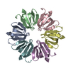
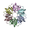
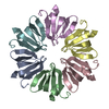
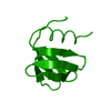
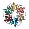
| Title | H57T Hfq from Pseudomonas aeruginosa | ||||||
 Components Components | Protein hfq | ||||||
 Keywords Keywords | RNA BINDING PROTEIN / Hfq / protein tertiary structure / RNA-binding / Stress response / RNA-BINDING PROTEIN | ||||||
| Function / homology |  Function and homology information Function and homology informationregulation of translation, ncRNA-mediated / regulation of carbon utilization / regulation of RNA stability / quorum sensing / regulation of translation / regulation of DNA-templated transcription / RNA binding / cytosol Similarity search - Function | ||||||
| Biological species |   Pseudomonas aeruginosa (bacteria) Pseudomonas aeruginosa (bacteria) | ||||||
| Method |  X-RAY DIFFRACTION / SYNCHROTRON / MOLECULAR REPLACEMENT / Resolution: 1.7 Å X-RAY DIFFRACTION / SYNCHROTRON / MOLECULAR REPLACEMENT / Resolution: 1.7 Å | ||||||
 Authors Authors | Moskaleva, O. / Melnik, B. / Gabdulkhakov, A. / Garber, M. / Nikonov, S. / Stolboushkina, E. / Nikulin, A. | ||||||
 Citation Citation | Journal: Acta Crystallogr.,Sect.F / Year: 2010 Title: The structures of mutant forms of Hfq from Pseudomonas aeruginosa reveal the importance of the conserved His57 for the protein hexamer organization. Authors: Moskaleva, O. / Melnik, B. / Gabdulkhakov, A. / Garber, M. / Nikonov, S. / Stolboushkina, E. / Nikulin, A. #1: Journal: Acta Crystallogr.,Sect.D / Year: 2005Title: Structure of Pseudomonas aeruginosa Hfq protein. Authors: Nikulin, A. / Stolboushkina, E. / Perederina, A. / Vassilieva, I. / Blaesi, U. / Moll, I. / Kachalova, G. / Yokoyama, S. / Vassylyev, D. / Garber, M. / Nikonov, S. | ||||||
| History |
|
- Structure visualization
| Structure viewer | Molecule: MolmilJmol/JSmol |
|---|
- Downloads & links
-Download
| PDBx/mmCIF format | 3inz.cif.gz | 197.8 KB | Display | PDBx/mmCIF format |
|---|---|---|---|---|
| PDB format | pdb3inz.ent.gz | 158.1 KB | Display | PDB format |
| PDBx/mmJSON format | 3inz.json.gz | Tree view | PDBx/mmJSON format | |
| Others |  Other downloads Other downloads |
-Validation report
| Arichive directory | https://data.pdbj.org/pub/pdb/validation_reports/in/3inzftp://data.pdbj.org/pub/pdb/validation_reports/in/3inz | HTTPS FTP |
|---|
-Related structure data
| Related structure data | 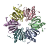 3m4gC 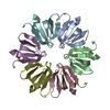 1u1sS S: Starting model for refinement C: citing same article ( |
|---|---|
| Similar structure data |


-Links
 PDBj
PDBj

- Assembly
| Deposited unit | 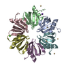
| ||||||||
|---|---|---|---|---|---|---|---|---|---|
| 1 |
| ||||||||
| Unit cell |
|
-Components
| #1: Protein | Mass: 9077.444 Da / Num. of mol.: 6 / Mutation: H57T Source method: isolated from a genetically manipulated source Source: (gene. exp.) Pseudomonas aeruginosa (bacteria) / Gene: hfq, PA4944 / Plasmid: pET22b / Production host: #2: Chemical | ChemComp-CD /   Mass: 112.411 Da / Num. of mol.: 9 / Source method: obtained synthetically / Formula: Cd Mass: 112.411 Da / Num. of mol.: 9 / Source method: obtained synthetically / Formula: Cd#3: Chemical | ChemComp-NA / |   Mass: 22.990 Da / Num. of mol.: 1 / Source method: obtained synthetically / Formula: Na Mass: 22.990 Da / Num. of mol.: 1 / Source method: obtained synthetically / Formula: Na#4: Chemical | ChemComp-CL / |   Mass: 35.453 Da / Num. of mol.: 1 / Source method: obtained synthetically / Formula: Cl Mass: 35.453 Da / Num. of mol.: 1 / Source method: obtained synthetically / Formula: Cl#5: Water | ChemComp-HOH / |  Mass: 18.015 Da / Num. of mol.: 379 / Source method: isolated from a natural source / Formula: H2O Mass: 18.015 Da / Num. of mol.: 379 / Source method: isolated from a natural source / Formula: H2O |
|---|
-Experimental details
-Experiment
| Experiment | Method: X-RAY DIFFRACTION / Number of used crystals: 1 |
|---|
- Sample preparation
| Crystal | Density Matthews: 2.09 Å3/Da / Density % sol: 41.15 % |
|---|---|
| Crystal grow | Temperature: 295 K / Method: vapor diffusion / pH: 8.5 Details: 4 mg/ml protein, 50 mM NaCl, 100 mM NH4Cl, 7,5% MMEPEG 2000, 50 mM Tris HCl, 10 mM CdCl2, pH 8.5, VAPOR DIFFUSION, temperature 295K |
-Data collection
| Diffraction | Mean temperature: 100 K |
|---|---|
| Diffraction source | Source: SYNCHROTRON / Site: EMBL/DESY, HAMBURG  / Beamline: X12 / Wavelength: 1 Å / Beamline: X12 / Wavelength: 1 Å |
| Detector | Type: MARMOSAIC 225 mm CCD / Detector: CCD / Date: Jun 26, 2006 |
| Radiation | Protocol: SINGLE WAVELENGTH / Monochromatic (M) / Laue (L): M / Scattering type: x-ray |
| Radiation wavelength | Wavelength: 1 Å / Relative weight: 1 |
| Reflection | Resolution: 1.7→30 Å / Num. obs: 50597 / % possible obs: 94.1 % / Observed criterion σ(F): 0 / Observed criterion σ(I): 0 / Redundancy: 6.7 % / Biso Wilson estimate: 31.2 Å2 / Rmerge(I) obs: 0.04 / Net I/σ(I): 27.9 |
| Reflection shell | Resolution: 1.7→1.74 Å / Redundancy: 3.5 % / Rmerge(I) obs: 0.362 / Mean I/σ(I) obs: 3.9 / Num. unique all: 2983 / % possible all: 80.4 |
- Processing
| Software |
| |||||||||||||||||||||||||||||||||||||||||||||||||||||||||||||||||||||||||||||||||||||||||||||||||||||||||||||||||||||||||||||||||||||
|---|---|---|---|---|---|---|---|---|---|---|---|---|---|---|---|---|---|---|---|---|---|---|---|---|---|---|---|---|---|---|---|---|---|---|---|---|---|---|---|---|---|---|---|---|---|---|---|---|---|---|---|---|---|---|---|---|---|---|---|---|---|---|---|---|---|---|---|---|---|---|---|---|---|---|---|---|---|---|---|---|---|---|---|---|---|---|---|---|---|---|---|---|---|---|---|---|---|---|---|---|---|---|---|---|---|---|---|---|---|---|---|---|---|---|---|---|---|---|---|---|---|---|---|---|---|---|---|---|---|---|---|---|---|---|
| Refinement | Method to determine structure: MOLECULAR REPLACEMENT Starting model: 1U1S Resolution: 1.7→30 Å / SU ML: 0.03 / Isotropic thermal model: anisotropic / Cross valid method: THROUGHOUT / σ(F): 1.99 / Stereochemistry target values: Engh & Huber
| |||||||||||||||||||||||||||||||||||||||||||||||||||||||||||||||||||||||||||||||||||||||||||||||||||||||||||||||||||||||||||||||||||||
| Solvent computation | Shrinkage radii: 0.9 Å / VDW probe radii: 1.11 Å / Solvent model: FLAT BULK SOLVENT MODEL / Bsol: 60.003 Å2 / ksol: 0.384 e/Å3 | |||||||||||||||||||||||||||||||||||||||||||||||||||||||||||||||||||||||||||||||||||||||||||||||||||||||||||||||||||||||||||||||||||||
| Displacement parameters | Biso mean: 1.1587 Å2
| |||||||||||||||||||||||||||||||||||||||||||||||||||||||||||||||||||||||||||||||||||||||||||||||||||||||||||||||||||||||||||||||||||||
| Refinement step | Cycle: LAST / Resolution: 1.7→30 Å
| |||||||||||||||||||||||||||||||||||||||||||||||||||||||||||||||||||||||||||||||||||||||||||||||||||||||||||||||||||||||||||||||||||||
| Refine LS restraints |
| |||||||||||||||||||||||||||||||||||||||||||||||||||||||||||||||||||||||||||||||||||||||||||||||||||||||||||||||||||||||||||||||||||||
| LS refinement shell |
|
