 Movie
Movie Controller

[English] 日本語
 Yorodumi
- PDB-1o22: Crystal structure of an orphan protein (TM0875) from Thermotoga m... -
+ Open data

- Basic information
| Entry | Database: PDB / ID: 1o22 | ||||||
|---|---|---|---|---|---|---|---|
| Title | Crystal structure of an orphan protein (TM0875) from Thermotoga maritima at 2.00 A resolution | ||||||
 Components Components | orphan protein TM0875 | ||||||
 Keywords Keywords | UNKNOWN FUNCTION / ORPHAN PROTEIN / STRUCTURAL GENOMICS / JOINT CENTER FOR STRUCTURAL GENOMICS / JCSG / PROTEIN STRUCTURE INITIATIVE / PSI | ||||||
| Function / homology | Orphan Protein Tm0875; Chain: A; / Hypothetical protein TM0875 / Domain of unknown function DUF3855 / Hypothetical protein TM0875 / Domain of Unknown Function with PDB structure (DUF3855) / Alpha-Beta Complex / Alpha Beta / DUF3855 domain-containing protein Function and homology information Function and homology information | ||||||
| Biological species |   Thermotoga maritima (bacteria) Thermotoga maritima (bacteria) | ||||||
| Method |  X-RAY DIFFRACTION / SYNCHROTRON / MAD / Resolution: 2 Å X-RAY DIFFRACTION / SYNCHROTRON / MAD / Resolution: 2 Å | ||||||
 Authors Authors | Joint Center for Structural Genomics (JCSG) | ||||||
 Citation Citation | Journal: Proteins / Year: 2004 Title: Crystal structure of an orphan protein (TM0875) from Thermotoga maritima at 2.00-A resolution reveals a new fold. Authors: Bakolitsa, C. / Schwarzenbacher, R. / McMullan, D. / Brinen, L.S. / Canaves, J.M. / Dai, X. / Deacon, A.M. / Elsliger, M.A. / Eshagi, S. / Floyd, R. / Godzik, A. / Grittini, C. / Grzechnik, ...Authors: Bakolitsa, C. / Schwarzenbacher, R. / McMullan, D. / Brinen, L.S. / Canaves, J.M. / Dai, X. / Deacon, A.M. / Elsliger, M.A. / Eshagi, S. / Floyd, R. / Godzik, A. / Grittini, C. / Grzechnik, S.K. / Jaroszewski, L. / Karlak, C. / Klock, H.E. / Koesema, E. / Kovarik, J.S. / Kreusch, A. / Kuhn, P. / Lesley, S.A. / McPhillips, T.M. / Miller, M.D. / Morse, A. / Moy, K. / Ouyang, J. / Page, R. / Quijano, K. / Robb, A. / Spraggon, G. / Stevens, R.C. / van den Bedem, H. / Velasquez, J. / Vincent, J. / von Delft, F. / Wang, X. / West, B. / Wolf, G. / Hodgson, K.O. / Wooley, J. / Wilson, I.A. | ||||||
| History |
|
- Structure visualization
| Structure viewer | Molecule: MolmilJmol/JSmol |
|---|
- Downloads & links
-Download
| PDBx/mmCIF format | 1o22.cif.gz | 48.3 KB | Display | PDBx/mmCIF format |
|---|---|---|---|---|
| PDB format | pdb1o22.ent.gz | 33.9 KB | Display | PDB format |
| PDBx/mmJSON format | 1o22.json.gz | Tree view | PDBx/mmJSON format | |
| Others |  Other downloads Other downloads |
-Validation report
| Arichive directory | https://data.pdbj.org/pub/pdb/validation_reports/o2/1o22ftp://data.pdbj.org/pub/pdb/validation_reports/o2/1o22 | HTTPS FTP |
|---|
-Related structure data
| Similar structure data | |
|---|---|
| Other databases |


-Links
 PDBj
PDBj- Assembly
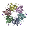
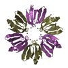
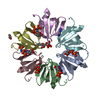
| Deposited unit | 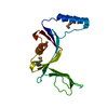
| ||||||||
|---|---|---|---|---|---|---|---|---|---|
| 1 | 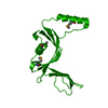
| ||||||||
| Unit cell |
|
-Components
| #1: Protein | Mass: 20343.340 Da / Num. of mol.: 1 Source method: isolated from a genetically manipulated source Source: (gene. exp.) Thermotoga maritima (bacteria) / Gene: TM0875 / Production host: |
|---|---|
| #2: Water | ChemComp-HOH /  Mass: 18.015 Da / Num. of mol.: 102 / Source method: isolated from a natural source / Formula: H2O Mass: 18.015 Da / Num. of mol.: 102 / Source method: isolated from a natural source / Formula: H2O |
| Has protein modification | Y |
-Experimental details
-Experiment
| Experiment | Method: X-RAY DIFFRACTION / Number of used crystals: 1 |
|---|
- Sample preparation
| Crystal | Density Matthews: 2.32 Å3/Da / Density % sol: 46.59 % | |||||||||||||||||||||||||||||||||||||||||||||||||||||||||||||||
|---|---|---|---|---|---|---|---|---|---|---|---|---|---|---|---|---|---|---|---|---|---|---|---|---|---|---|---|---|---|---|---|---|---|---|---|---|---|---|---|---|---|---|---|---|---|---|---|---|---|---|---|---|---|---|---|---|---|---|---|---|---|---|---|---|
| Crystal grow | Temperature: 293 K / pH: 8.5 Details: 25.5 % PEG 4000; 0.085 M Tris pH 8.5; 0.17 M Na(Ac), 15 % Glycerol, VAPOR DIFFUSION,SITTING DROP,NANODROP, temperature 293K, pH 8.50 | |||||||||||||||||||||||||||||||||||||||||||||||||||||||||||||||
| Crystal grow | *PLUS pH: 7.9 / Method: vapor diffusion | |||||||||||||||||||||||||||||||||||||||||||||||||||||||||||||||
| Components of the solutions | *PLUS
|
-Data collection
| Diffraction | Mean temperature: 100 K | ||||||||||||
|---|---|---|---|---|---|---|---|---|---|---|---|---|---|
| Diffraction source | Source: SYNCHROTRON / Site: ALS  / Beamline: 5.0.2 / Wavelength: 0.97780, 0.91840, 0.97920 / Beamline: 5.0.2 / Wavelength: 0.97780, 0.91840, 0.97920 | ||||||||||||
| Detector | Type: ADSC QUANTUM / Detector: CCD / Date: Oct 13, 2002 | ||||||||||||
| Radiation | Monochromator: DOUBLE-CRYSTAL SI(111) / Protocol: MAD / Monochromatic (M) / Laue (L): M / Scattering type: x-ray | ||||||||||||
| Radiation wavelength |
| ||||||||||||
| Reflection | Resolution: 2→32.159 Å / Num. obs: 12462 / % possible obs: 99 % / Redundancy: 11.8 % / Biso Wilson estimate: 36.49 Å2 / Rsym value: 0.061 / Net I/σ(I): 28.1 | ||||||||||||
| Reflection shell | Resolution: 2→2.05 Å / Redundancy: 6.2 % / Mean I/σ(I) obs: 3.2 / Rsym value: 0.375 / % possible all: 90.9 | ||||||||||||
| Reflection | *PLUS Highest resolution: 2 Å / Lowest resolution: 32.16 Å / Num. measured all: 148070 / Rmerge(I) obs: 0.061 | ||||||||||||
| Reflection shell | *PLUS % possible obs: 90.9 % / Rmerge(I) obs: 0.375 / Mean I/σ(I) obs: 2 |
- Processing
| Software |
| |||||||||||||||||||||||||||||||||||||||||||||||||||||||||||||||||||||||||||||||||||||||||||||||||||||||||||||||||||||||||||||||||||||||||||||||||||||||||||
|---|---|---|---|---|---|---|---|---|---|---|---|---|---|---|---|---|---|---|---|---|---|---|---|---|---|---|---|---|---|---|---|---|---|---|---|---|---|---|---|---|---|---|---|---|---|---|---|---|---|---|---|---|---|---|---|---|---|---|---|---|---|---|---|---|---|---|---|---|---|---|---|---|---|---|---|---|---|---|---|---|---|---|---|---|---|---|---|---|---|---|---|---|---|---|---|---|---|---|---|---|---|---|---|---|---|---|---|---|---|---|---|---|---|---|---|---|---|---|---|---|---|---|---|---|---|---|---|---|---|---|---|---|---|---|---|---|---|---|---|---|---|---|---|---|---|---|---|---|---|---|---|---|---|---|---|---|
| Refinement | Method to determine structure: MAD / Resolution: 2→29.49 Å / Cor.coef. Fo:Fc: 0.954 / Cor.coef. Fo:Fc free: 0.933 / SU B: 8.183 / SU ML: 0.111 / TLS residual ADP flag: LIKELY RESIDUAL / Isotropic thermal model: Isotropic / Cross valid method: THROUGHOUT / σ(F): 0 / ESU R: 0.175 / ESU R Free: 0.167 / Stereochemistry target values: MAXIMUM LIKELIHOOD Details: THE BIOLOGICAL UNIT APPEARS TO BE A DIMER THAT STRADDLES THE CRYSTALLOGRAPHIC 4-FOLD. TLS GROUPS WERE CHOSEN WITH REFERENCE TO THE DIMER. WEAK DENSITY BOTH BEFORE FIRST AND AFTER LAST ...Details: THE BIOLOGICAL UNIT APPEARS TO BE A DIMER THAT STRADDLES THE CRYSTALLOGRAPHIC 4-FOLD. TLS GROUPS WERE CHOSEN WITH REFERENCE TO THE DIMER. WEAK DENSITY BOTH BEFORE FIRST AND AFTER LAST RESIDUE SUGGESTS THE GENERAL POSITION OF MORE N- AND C-TERMINAL RESIDUES THAN MODELLED HERE. CNS_SOLVE 1.1/CNS/XFIT/CCP4/TLS WERE ALSO USED IN REFINEMENT.
| |||||||||||||||||||||||||||||||||||||||||||||||||||||||||||||||||||||||||||||||||||||||||||||||||||||||||||||||||||||||||||||||||||||||||||||||||||||||||||
| Solvent computation | Ion probe radii: 0.8 Å / Shrinkage radii: 0.8 Å / VDW probe radii: 1.4 Å / Solvent model: BABINET MODEL WITH MASK | |||||||||||||||||||||||||||||||||||||||||||||||||||||||||||||||||||||||||||||||||||||||||||||||||||||||||||||||||||||||||||||||||||||||||||||||||||||||||||
| Displacement parameters | Biso mean: 23.83 Å2
| |||||||||||||||||||||||||||||||||||||||||||||||||||||||||||||||||||||||||||||||||||||||||||||||||||||||||||||||||||||||||||||||||||||||||||||||||||||||||||
| Refinement step | Cycle: LAST / Resolution: 2→29.49 Å
| |||||||||||||||||||||||||||||||||||||||||||||||||||||||||||||||||||||||||||||||||||||||||||||||||||||||||||||||||||||||||||||||||||||||||||||||||||||||||||
| Refine LS restraints |
| |||||||||||||||||||||||||||||||||||||||||||||||||||||||||||||||||||||||||||||||||||||||||||||||||||||||||||||||||||||||||||||||||||||||||||||||||||||||||||
| LS refinement shell | Resolution: 2→2.05 Å / Total num. of bins used: 20 /
| |||||||||||||||||||||||||||||||||||||||||||||||||||||||||||||||||||||||||||||||||||||||||||||||||||||||||||||||||||||||||||||||||||||||||||||||||||||||||||
| Refinement TLS params. | Method: refined / Refine-ID: X-RAY DIFFRACTION
| |||||||||||||||||||||||||||||||||||||||||||||||||||||||||||||||||||||||||||||||||||||||||||||||||||||||||||||||||||||||||||||||||||||||||||||||||||||||||||
| Refinement TLS group |
| |||||||||||||||||||||||||||||||||||||||||||||||||||||||||||||||||||||||||||||||||||||||||||||||||||||||||||||||||||||||||||||||||||||||||||||||||||||||||||
| Software | *PLUS Name: REFMAC / Version: 5.1.995 / Classification: refinement | |||||||||||||||||||||||||||||||||||||||||||||||||||||||||||||||||||||||||||||||||||||||||||||||||||||||||||||||||||||||||||||||||||||||||||||||||||||||||||
| Refinement | *PLUS Lowest resolution: 32.16 Å / Num. reflection obs: 12461 / % reflection Rfree: 7.1 % | |||||||||||||||||||||||||||||||||||||||||||||||||||||||||||||||||||||||||||||||||||||||||||||||||||||||||||||||||||||||||||||||||||||||||||||||||||||||||||
| Solvent computation | *PLUS | |||||||||||||||||||||||||||||||||||||||||||||||||||||||||||||||||||||||||||||||||||||||||||||||||||||||||||||||||||||||||||||||||||||||||||||||||||||||||||
| Displacement parameters | *PLUS | |||||||||||||||||||||||||||||||||||||||||||||||||||||||||||||||||||||||||||||||||||||||||||||||||||||||||||||||||||||||||||||||||||||||||||||||||||||||||||
| Refine LS restraints | *PLUS
|
