 Movie
Movie Controller


+ Open data

- Basic information
| Entry | Database: PDB / ID: 4jri | ||||||
|---|---|---|---|---|---|---|---|
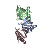
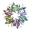
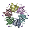
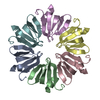
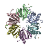
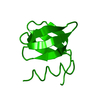
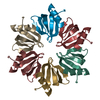
| Title | Crystal Structure of Escherichia coli Hfq Proximal Edge Mutant | ||||||
 Components Components | Protein hfq | ||||||
 Keywords Keywords | RNA BINDING PROTEIN / Riboregulator / Post-transcriptional regulator | ||||||
| Function / homology |  Function and homology information Function and homology informationsRNA-mediated post-transcriptional gene silencing / positive regulation of translation, ncRNA-mediated / bacterial nucleoid / regulation of translation, ncRNA-mediated / bent DNA binding / regulation of RNA stability / RNA folding chaperone / tRNA processing / tRNA binding / regulation of DNA-templated transcription ...sRNA-mediated post-transcriptional gene silencing / positive regulation of translation, ncRNA-mediated / bacterial nucleoid / regulation of translation, ncRNA-mediated / bent DNA binding / regulation of RNA stability / RNA folding chaperone / tRNA processing / tRNA binding / regulation of DNA-templated transcription / DNA binding / RNA binding / ATP binding / cytosol Similarity search - Function | ||||||
| Biological species |  | ||||||
| Method |  X-RAY DIFFRACTION / SYNCHROTRON / MOLECULAR REPLACEMENT / Resolution: 1.829 Å X-RAY DIFFRACTION / SYNCHROTRON / MOLECULAR REPLACEMENT / Resolution: 1.829 Å | ||||||
 Authors Authors | Robinson, K.E. / Orans, J. | ||||||
 Citation Citation | Journal: Nucleic Acids Res. / Year: 2014 Title: Mapping Hfq-RNA interaction surfaces using tryptophan fluorescence quenching. Authors: Robinson, K.E. / Orans, J. / Kovach, A.R. / Link, T.M. / Brennan, R.G. | ||||||
| History |
|
- Structure visualization
| Structure viewer | Molecule: MolmilJmol/JSmol |
|---|
- Downloads & links
-Download
| PDBx/mmCIF format | 4jri.cif.gz | 66.2 KB | Display | PDBx/mmCIF format |
|---|---|---|---|---|
| PDB format | pdb4jri.ent.gz | 50.6 KB | Display | PDB format |
| PDBx/mmJSON format | 4jri.json.gz | Tree view | PDBx/mmJSON format | |
| Others |  Other downloads Other downloads |
-Validation report
| Arichive directory | https://data.pdbj.org/pub/pdb/validation_reports/jr/4jriftp://data.pdbj.org/pub/pdb/validation_reports/jr/4jri | HTTPS FTP |
|---|
-Related structure data


-Links
 PDBj
PDBj

- Assembly
| Deposited unit | 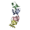
| |||||||||||||||
|---|---|---|---|---|---|---|---|---|---|---|---|---|---|---|---|---|
| 1 | 
| |||||||||||||||
| 2 | 
| |||||||||||||||
| Unit cell |
| |||||||||||||||
| Components on special symmetry positions |
|
-Components
| #1: Protein | Mass: 7673.911 Da / Num. of mol.: 4 / Fragment: UNP residues 2-69 / Mutation: F39W Source method: isolated from a genetically manipulated source Source: (gene. exp.) #2: Water | ChemComp-HOH / |  Mass: 18.015 Da / Num. of mol.: 128 / Source method: isolated from a natural source / Formula: H2O Mass: 18.015 Da / Num. of mol.: 128 / Source method: isolated from a natural source / Formula: H2O |
|---|
-Experimental details
-Experiment
| Experiment | Method: X-RAY DIFFRACTION / Number of used crystals: 1 |
|---|
- Sample preparation
| Crystal | Density Matthews: 1.98 Å3/Da / Density % sol: 38.03 % |
|---|
-Data collection
| Diffraction source | Source: SYNCHROTRON / Site: APS  / Beamline: 22-BM / Beamline: 22-BM |
|---|---|
| Detector | Type: MARMOSAIC 225 mm CCD / Detector: CCD |
| Radiation | Protocol: SINGLE WAVELENGTH / Monochromatic (M) / Laue (L): M / Scattering type: x-ray |
| Radiation wavelength | Relative weight: 1 |
| Reflection | Resolution: 1.829→24.077 Å / Num. obs: 21700 / Rsym value: 0.06 |
- Processing
| Software | Name: PHENIX / Version: (phenix.refine: 1.8_1069) / Classification: refinement | |||||||||||||||||||||||||||||||||||||||||||||||||||||||||||||||
|---|---|---|---|---|---|---|---|---|---|---|---|---|---|---|---|---|---|---|---|---|---|---|---|---|---|---|---|---|---|---|---|---|---|---|---|---|---|---|---|---|---|---|---|---|---|---|---|---|---|---|---|---|---|---|---|---|---|---|---|---|---|---|---|---|
| Refinement | Method to determine structure: MOLECULAR REPLACEMENT / Resolution: 1.829→24.077 Å / SU ML: 0.24 / σ(F): 1.34 / Phase error: 26.6 / Stereochemistry target values: MLHL
| |||||||||||||||||||||||||||||||||||||||||||||||||||||||||||||||
| Solvent computation | Shrinkage radii: 0.9 Å / VDW probe radii: 1.11 Å / Solvent model: FLAT BULK SOLVENT MODEL | |||||||||||||||||||||||||||||||||||||||||||||||||||||||||||||||
| Refinement step | Cycle: LAST / Resolution: 1.829→24.077 Å
| |||||||||||||||||||||||||||||||||||||||||||||||||||||||||||||||
| Refine LS restraints |
| |||||||||||||||||||||||||||||||||||||||||||||||||||||||||||||||
| LS refinement shell |
|
