 Movie
Movie Controller

[English] 日本語
 Yorodumi
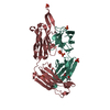
- PDB-3dsf: Crystal structure of anti-osteopontin antibody 23C3 in complex wi... -
+ Open data

- Basic information
| Entry | Database: PDB / ID: 3dsf | |||||||||
|---|---|---|---|---|---|---|---|---|---|---|
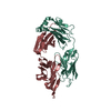
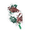
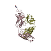
| Title | Crystal structure of anti-osteopontin antibody 23C3 in complex with W43A mutated epitope peptide | |||||||||
 Components Components |
| |||||||||
 Keywords Keywords | IMMUNE SYSTEM / Fab / osteopontin / OPN / antibody-antigen complex | |||||||||
| Function / homology |  Function and homology information Function and homology informationnegative regulation of collateral sprouting of intact axon in response to injury / positive regulation of estradiol secretion / androgen catabolic process / biomineral tissue development / response to macrophage colony-stimulating factor / response to 2,3,7,8-tetrachlorodibenzodioxine / cellular response to testosterone stimulus / Signaling by PDGF / RUNX3 Regulates Immune Response and Cell Migration / positive regulation of bone resorption ...negative regulation of collateral sprouting of intact axon in response to injury / positive regulation of estradiol secretion / androgen catabolic process / biomineral tissue development / response to macrophage colony-stimulating factor / response to 2,3,7,8-tetrachlorodibenzodioxine / cellular response to testosterone stimulus / Signaling by PDGF / RUNX3 Regulates Immune Response and Cell Migration / positive regulation of bone resorption / response to vitamin D / extracellular matrix binding / response to steroid hormone / Mechanical load activates signaling by PIEZO1 and integrins in osteocytes / decidualization / small molecule binding / Integrin cell surface interactions / embryo implantation / Degradation of the extracellular matrix / cell projection / cytokine activity / Post-translational protein phosphorylation / integrin binding / Regulation of Insulin-like Growth Factor (IGF) transport and uptake by Insulin-like Growth Factor Binding Proteins (IGFBPs) / osteoblast differentiation / cell adhesion / endoplasmic reticulum lumen / calcium ion binding / positive regulation of DNA-templated transcription / perinuclear region of cytoplasm / Golgi apparatus / : / extracellular exosome / extracellular region Similarity search - Function | |||||||||
| Biological species |  | |||||||||
| Method |  X-RAY DIFFRACTION / MOLECULAR REPLACEMENT / Resolution: 2.8 Å X-RAY DIFFRACTION / MOLECULAR REPLACEMENT / Resolution: 2.8 Å | |||||||||
 Authors Authors | Du, J. / Zhong, C. / Yang, H. / Ding, J. | |||||||||
 Citation Citation | Journal: J.Mol.Biol. / Year: 2008 Title: Molecular basis of recognition of human osteopontin by 23C3, a potential therapeutic antibody for treatment of rheumatoid arthritis Authors: Du, J. / Hou, S. / Zhong, C. / Lai, Z. / Yang, H. / Dai, J. / Zhang, D. / Wang, H. / Guo, Y. / Ding, J. | |||||||||
| History |
|
- Structure visualization
| Structure viewer | Molecule: MolmilJmol/JSmol |
|---|
- Downloads & links
-Download
| PDBx/mmCIF format | 3dsf.cif.gz | 164.9 KB | Display | PDBx/mmCIF format |
|---|---|---|---|---|
| PDB format | pdb3dsf.ent.gz | 129.3 KB | Display | PDB format |
| PDBx/mmJSON format | 3dsf.json.gz | Tree view | PDBx/mmJSON format | |
| Others |  Other downloads Other downloads |
-Validation report
| Arichive directory | https://data.pdbj.org/pub/pdb/validation_reports/ds/3dsfftp://data.pdbj.org/pub/pdb/validation_reports/ds/3dsf | HTTPS FTP |
|---|
-Related structure data
| Related structure data | 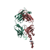 3cxdSC S: Starting model for refinement C: citing same article ( |
|---|---|
| Similar structure data |


-Links
 PDBj
PDBj


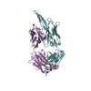
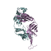
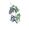
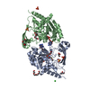
- Assembly
| Deposited unit | 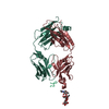
| ||||||||
|---|---|---|---|---|---|---|---|---|---|
| 1 |
| ||||||||
| Unit cell |
|
-Components
| #1: Antibody | Mass: 23070.371 Da / Num. of mol.: 1 / Source method: isolated from a natural source / Source: (natural) |
|---|---|
| #2: Antibody | Mass: 23131.977 Da / Num. of mol.: 1 / Source method: isolated from a natural source / Source: (natural) |
| #3: Protein/peptide | Mass: 1241.370 Da / Num. of mol.: 1 / Source method: obtained synthetically Details: the peptide VATALNPDPSQK is synthesized at Shanghai HD Bioscience Company References: UniProt: P10451 |
| #4: Polysaccharide | alpha-D-mannopyranose-(1-3)-beta-D-mannopyranose-(1-4)-2-acetamido-2-deoxy-beta-D-glucopyranose-(1- ...alpha-D-mannopyranose-(1-3)-beta-D-mannopyranose-(1-4)-2-acetamido-2-deoxy-beta-D-glucopyranose-(1-4)-2-acetamido-2-deoxy-beta-D-glucopyranose Source method: isolated from a genetically manipulated source |
| Has protein modification | Y |
-Experimental details
-Experiment
| Experiment | Method: X-RAY DIFFRACTION / Number of used crystals: 1 |
|---|
- Sample preparation
| Crystal | Density Matthews: 2.47 Å3/Da / Density % sol: 50.24 % |
|---|---|
| Crystal grow | Temperature: 277 K / Method: vapor diffusion, hanging drop / pH: 7.9 Details: 0.2M di-ammonium hydrogen phosphate, 20% PEG3350, pH 7.9, VAPOR DIFFUSION, HANGING DROP, temperature 277K |
-Data collection
| Diffraction | Mean temperature: 100 K |
|---|---|
| Diffraction source | Source: ROTATING ANODE / Type: RIGAKU / Wavelength: 1.5418 Å |
| Detector | Type: RIGAKU RAXIS IV++ / Detector: IMAGE PLATE / Date: Jul 1, 2008 |
| Radiation | Protocol: SINGLE WAVELENGTH / Monochromatic (M) / Laue (L): M / Scattering type: x-ray |
| Radiation wavelength | Wavelength: 1.5418 Å / Relative weight: 1 |
| Reflection | Resolution: 2.8→20 Å / Num. all: 12159 / Num. obs: 12086 / % possible obs: 99.4 % / Rmerge(I) obs: 0.084 |
| Reflection shell | Resolution: 2.8→2.9 Å / Rmerge(I) obs: 0.229 / Mean I/σ(I) obs: 3.1 / Rsym value: 0.229 / % possible all: 99.4 |
- Processing
| Software |
| ||||||||||||||||||||||||||||||||||||||||||||||||||||||||||||||||||||||||||||||||||||||||||||||||||||
|---|---|---|---|---|---|---|---|---|---|---|---|---|---|---|---|---|---|---|---|---|---|---|---|---|---|---|---|---|---|---|---|---|---|---|---|---|---|---|---|---|---|---|---|---|---|---|---|---|---|---|---|---|---|---|---|---|---|---|---|---|---|---|---|---|---|---|---|---|---|---|---|---|---|---|---|---|---|---|---|---|---|---|---|---|---|---|---|---|---|---|---|---|---|---|---|---|---|---|---|---|---|
| Refinement | Method to determine structure: MOLECULAR REPLACEMENT Starting model: PDB ENTRY 3CXD Resolution: 2.8→20 Å / Cor.coef. Fo:Fc: 0.885 / Cor.coef. Fo:Fc free: 0.83 / Cross valid method: THROUGHOUT / σ(F): 0 / σ(I): 0 / ESU R Free: 0.459 / Stereochemistry target values: MAXIMUM LIKELIHOOD
| ||||||||||||||||||||||||||||||||||||||||||||||||||||||||||||||||||||||||||||||||||||||||||||||||||||
| Solvent computation | Ion probe radii: 0.8 Å / Shrinkage radii: 0.8 Å / VDW probe radii: 1.2 Å / Solvent model: MASK | ||||||||||||||||||||||||||||||||||||||||||||||||||||||||||||||||||||||||||||||||||||||||||||||||||||
| Displacement parameters | Biso mean: 33.454 Å2
| ||||||||||||||||||||||||||||||||||||||||||||||||||||||||||||||||||||||||||||||||||||||||||||||||||||
| Refinement step | Cycle: LAST / Resolution: 2.8→20 Å
| ||||||||||||||||||||||||||||||||||||||||||||||||||||||||||||||||||||||||||||||||||||||||||||||||||||
| Refine LS restraints |
| ||||||||||||||||||||||||||||||||||||||||||||||||||||||||||||||||||||||||||||||||||||||||||||||||||||
| LS refinement shell | Resolution: 2.8→2.871 Å / Total num. of bins used: 20
|
