 Movie
Movie Controller

[English] 日本語
 Yorodumi
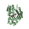
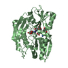
- PDB-3cvl: Structure of Peroxisomal Targeting Signal 1 (PTS1) binding domain... -
+ Open data

- Basic information
| Entry | Database: PDB / ID: 3cvl | ||||||
|---|---|---|---|---|---|---|---|
| Title | Structure of Peroxisomal Targeting Signal 1 (PTS1) binding domain of Trypanosoma brucei Peroxin 5 (TbPEX5)complexed to T. brucei Phosphofructokinase (PFK) PTS1 peptide | ||||||
 Components Components |
| ||||||
 Keywords Keywords | TRANSPORT PROTEIN / TPR motifs / TPR protein / Peroxin 5 / PEX5 / PTS1 binding domain / Protein-peptide complex / Receptor / TPR repeat | ||||||
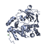
| Function / homology |  Function and homology information Function and homology informationperoxisome matrix targeting signal-1 binding / 6-phosphofructokinase / protein import into peroxisome matrix, docking / 6-phosphofructokinase activity / glycosome / fructose 6-phosphate metabolic process / peroxisomal membrane / phosphate ion binding / glycolytic process / ATP binding ...peroxisome matrix targeting signal-1 binding / 6-phosphofructokinase / protein import into peroxisome matrix, docking / 6-phosphofructokinase activity / glycosome / fructose 6-phosphate metabolic process / peroxisomal membrane / phosphate ion binding / glycolytic process / ATP binding / metal ion binding / cytosol Similarity search - Function | ||||||
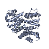
| Biological species |  | ||||||
| Method |  X-RAY DIFFRACTION / MOLECULAR REPLACEMENT / Resolution: 2.15 Å X-RAY DIFFRACTION / MOLECULAR REPLACEMENT / Resolution: 2.15 Å | ||||||
 Authors Authors | Sampathkumar, P. / Roach, C. / Michels, P.A.M. / Hol, W.G.J. | ||||||
 Citation Citation | Journal: J.Mol.Biol. / Year: 2008 Title: Structural Insights into the recognition of peroxisomal targeting signal 1 by Trypanosoma brucei peroxin 5. Authors: Sampathkumar, P. / Roach, C. / Michels, P.A. / Hol, W.G. | ||||||
| History |
|
- Structure visualization
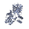
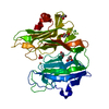
| Structure viewer | Molecule: MolmilJmol/JSmol |
|---|
- Downloads & links
-Download
| PDBx/mmCIF format | 3cvl.cif.gz | 72.9 KB | Display | PDBx/mmCIF format |
|---|---|---|---|---|
| PDB format | pdb3cvl.ent.gz | 52.9 KB | Display | PDB format |
| PDBx/mmJSON format | 3cvl.json.gz | Tree view | PDBx/mmJSON format | |
| Others |  Other downloads Other downloads |
-Validation report
| Arichive directory | https://data.pdbj.org/pub/pdb/validation_reports/cv/3cvlftp://data.pdbj.org/pub/pdb/validation_reports/cv/3cvl | HTTPS FTP |
|---|
-Related structure data
| Related structure data |  3cv0C  3cvnC  3cvpC  3cvqC  1fchS C: citing same article ( S: Starting model for refinement |
|---|---|
| Similar structure data |

-Links
 PDBj
PDBj


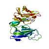
- Assembly
| Deposited unit | 
| ||||||||
|---|---|---|---|---|---|---|---|---|---|
| 1 |
| ||||||||
| Unit cell |
| ||||||||
| Details | THE BIOLOGICAL UNIT OF PROTEIN PEX5 IS MONOMER |
-Components
| #1: Protein | Mass: 36545.844 Da / Num. of mol.: 1 / Fragment: Binding domain,UNP residues 332-655 / Mutation: K378A/E379A Source method: isolated from a genetically manipulated source Source: (gene. exp.)  |
|---|---|
| #2: Protein/peptide | Mass: 840.963 Da / Num. of mol.: 1 / Source method: obtained synthetically / References: UniProt: O15648 |
| #3: Water | ChemComp-HOH /  Mass: 18.015 Da / Num. of mol.: 67 / Source method: isolated from a natural source / Formula: H2O Mass: 18.015 Da / Num. of mol.: 67 / Source method: isolated from a natural source / Formula: H2O |
-Experimental details
-Experiment
| Experiment | Method: X-RAY DIFFRACTION / Number of used crystals: 1 |
|---|
- Sample preparation
| Crystal | Density Matthews: 1.89 Å3/Da / Density % sol: 34.9 % |
|---|---|
| Crystal grow | Temperature: 298 K / Method: vapor diffusion, sitting drop / pH: 4.8 Details: 2.3M Potassium acetate, 0.1M sodium citrate monohydrate, pH 4.8, VAPOR DIFFUSION, SITTING DROP, temperature 298K |
-Data collection
| Diffraction | Mean temperature: 100 K |
|---|---|
| Diffraction source | Source: ROTATING ANODE / Type: RIGAKU MICROMAX-007 HF / Wavelength: 1.5418 |
| Detector | Type: RIGAKU SATURN 944 / Detector: CCD / Date: Nov 15, 2006 / Details: Osmic VariMax optics |
| Radiation | Protocol: SINGLE WAVELENGTH / Monochromatic (M) / Laue (L): M / Scattering type: x-ray |
| Radiation wavelength | Wavelength: 1.5418 Å / Relative weight: 1 |
| Reflection | Resolution: 2.08→26.4 Å / Num. all: 16165 / Num. obs: 16165 / % possible obs: 97.6 % / Redundancy: 3.27 % / Biso Wilson estimate: 25.52 Å2 / Rsym value: 0.096 / Net I/σ(I): 6.5 |
| Reflection shell | Resolution: 2.08→2.15 Å / Redundancy: 2.73 % / Mean I/σ(I) obs: 2.1 / Rsym value: 0.317 / % possible all: 85.4 |
- Processing
| Software |
| |||||||||||||||||||||||||||||||||||||||||||||||||||||||||||||||||||||||||||||||||||||||||||||||||||||||||||||||||||||||||||||
|---|---|---|---|---|---|---|---|---|---|---|---|---|---|---|---|---|---|---|---|---|---|---|---|---|---|---|---|---|---|---|---|---|---|---|---|---|---|---|---|---|---|---|---|---|---|---|---|---|---|---|---|---|---|---|---|---|---|---|---|---|---|---|---|---|---|---|---|---|---|---|---|---|---|---|---|---|---|---|---|---|---|---|---|---|---|---|---|---|---|---|---|---|---|---|---|---|---|---|---|---|---|---|---|---|---|---|---|---|---|---|---|---|---|---|---|---|---|---|---|---|---|---|---|---|---|---|
| Refinement | Method to determine structure: MOLECULAR REPLACEMENT Starting model: PDB code 1FCH Resolution: 2.15→26.17 Å / Cor.coef. Fo:Fc: 0.937 / Cor.coef. Fo:Fc free: 0.912 / SU B: 7.911 / SU ML: 0.206 / Cross valid method: THROUGHOUT / σ(F): 0 / ESU R: 0.341 / ESU R Free: 0.253 Stereochemistry target values: MAXIMUM LIKELIHOOD WITH PHASES
| |||||||||||||||||||||||||||||||||||||||||||||||||||||||||||||||||||||||||||||||||||||||||||||||||||||||||||||||||||||||||||||
| Solvent computation | Ion probe radii: 0.8 Å / Shrinkage radii: 0.8 Å / VDW probe radii: 1.4 Å / Solvent model: BABINET MODEL WITH MASK | |||||||||||||||||||||||||||||||||||||||||||||||||||||||||||||||||||||||||||||||||||||||||||||||||||||||||||||||||||||||||||||
| Displacement parameters | Biso mean: 23.242 Å2
| |||||||||||||||||||||||||||||||||||||||||||||||||||||||||||||||||||||||||||||||||||||||||||||||||||||||||||||||||||||||||||||
| Refinement step | Cycle: LAST / Resolution: 2.15→26.17 Å
| |||||||||||||||||||||||||||||||||||||||||||||||||||||||||||||||||||||||||||||||||||||||||||||||||||||||||||||||||||||||||||||
| Refine LS restraints |
| |||||||||||||||||||||||||||||||||||||||||||||||||||||||||||||||||||||||||||||||||||||||||||||||||||||||||||||||||||||||||||||
| LS refinement shell | Resolution: 2.15→2.206 Å / Total num. of bins used: 20
|
