 Movie
Movie Controller

[English] 日本語
 Yorodumi
- PDB-3cv0: Structure of Peroxisomal Targeting Signal 1 (PTS1) binding domain... -
+ Open data

- Basic information
| Entry | Database: PDB / ID: 3cv0 | ||||||
|---|---|---|---|---|---|---|---|
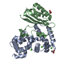
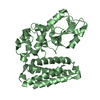
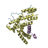
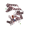
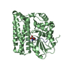
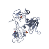
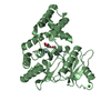
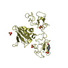
| Title | Structure of Peroxisomal Targeting Signal 1 (PTS1) binding domain of Trypanosoma brucei Peroxin 5 (TbPEX5)complexed to T. brucei Phosphoglucoisomerase (PGI) PTS1 peptide | ||||||
 Components Components |
| ||||||
 Keywords Keywords | TRANSPORT PROTEIN / TPR motifs / TPR protein / Peroxin 5 / PEX5 / PTS1 binding domain / Protein-peptide complex / Receptor / TPR repeat | ||||||
| Function / homology |  Function and homology information Function and homology informationperoxisome matrix targeting signal-1 binding / protein import into peroxisome matrix, docking / glucose-6-phosphate isomerase / glucose-6-phosphate isomerase activity / glucose 6-phosphate metabolic process / glycosome / carbohydrate derivative binding / monosaccharide binding / peroxisomal membrane / glycolytic process ...peroxisome matrix targeting signal-1 binding / protein import into peroxisome matrix, docking / glucose-6-phosphate isomerase / glucose-6-phosphate isomerase activity / glucose 6-phosphate metabolic process / glycosome / carbohydrate derivative binding / monosaccharide binding / peroxisomal membrane / glycolytic process / gluconeogenesis / metal ion binding / cytoplasm / cytosol Similarity search - Function | ||||||
| Biological species |  | ||||||
| Method |  X-RAY DIFFRACTION / MOLECULAR REPLACEMENT / Resolution: 2 Å X-RAY DIFFRACTION / MOLECULAR REPLACEMENT / Resolution: 2 Å | ||||||
 Authors Authors | Sampathkumar, P. / Roach, C. / Michels, P.A.M. / Hol, W.G.J. | ||||||
 Citation Citation | Journal: J.Mol.Biol. / Year: 2008 Title: Structural Insights into the recognition of peroxisomal targeting signal 1 by Trypanosoma brucei peroxin 5. Authors: Sampathkumar, P. / Roach, C. / Michels, P.A. / Hol, W.G. | ||||||
| History |
|
- Structure visualization
| Structure viewer | Molecule: MolmilJmol/JSmol |
|---|
- Downloads & links
-Download
| PDBx/mmCIF format | 3cv0.cif.gz | 77.2 KB | Display | PDBx/mmCIF format |
|---|---|---|---|---|
| PDB format | pdb3cv0.ent.gz | 56.9 KB | Display | PDB format |
| PDBx/mmJSON format | 3cv0.json.gz | Tree view | PDBx/mmJSON format | |
| Others |  Other downloads Other downloads |
-Validation report
| Arichive directory | https://data.pdbj.org/pub/pdb/validation_reports/cv/3cv0ftp://data.pdbj.org/pub/pdb/validation_reports/cv/3cv0 | HTTPS FTP |
|---|
-Related structure data
| Related structure data |  3cvlC 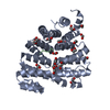 3cvnC  3cvpC 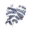 3cvqC  1fhcS C: citing same article ( S: Starting model for refinement |
|---|---|
| Similar structure data |


-Links
 PDBj
PDBj


- Assembly
| Deposited unit | 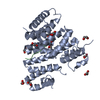
| ||||||||
|---|---|---|---|---|---|---|---|---|---|
| 1 |
| ||||||||
| Unit cell |
| ||||||||
| Details | THE BIOLOGICAL UNIT OF PROTEIN PEX5 IS MONOMER |
-Components
| #1: Protein | Mass: 36545.844 Da / Num. of mol.: 1 / Fragment: UNP residues 332-655 / Mutation: K378A/E379A Source method: isolated from a genetically manipulated source Source: (gene. exp.)  | ||||
|---|---|---|---|---|---|
| #2: Protein/peptide | Mass: 859.946 Da / Num. of mol.: 1 / Source method: obtained synthetically / References: UniProt: P13377 | ||||
| #3: Chemical | ChemComp-EDO /   Mass: 62.068 Da / Num. of mol.: 12 / Source method: obtained synthetically / Formula: C2H6O2 Mass: 62.068 Da / Num. of mol.: 12 / Source method: obtained synthetically / Formula: C2H6O2#4: Water | ChemComp-HOH / |  Mass: 18.015 Da / Num. of mol.: 116 / Source method: isolated from a natural source / Formula: H2O Mass: 18.015 Da / Num. of mol.: 116 / Source method: isolated from a natural source / Formula: H2OSequence details | ACE IS COVALENTLY | |
-Experimental details
-Experiment
| Experiment | Method: X-RAY DIFFRACTION / Number of used crystals: 1 |
|---|
- Sample preparation
| Crystal | Density Matthews: 2.04 Å3/Da / Density % sol: 39.83 % |
|---|---|
| Crystal grow | Temperature: 298 K / Method: vapor diffusion, sitting drop / pH: 4.8 Details: 2.3M Potassium acetate, 0.1M sodium citrate monohydrate, pH 4.8, vapor diffusion, sitting drop, temperature 298K |
-Data collection
| Diffraction | Mean temperature: 100 K |
|---|---|
| Diffraction source | Source: ROTATING ANODE / Type: RIGAKU MICROMAX-007 HF / Wavelength: 1.5418 |
| Detector | Type: RIGAKU SATURN 944 / Detector: CCD / Date: Dec 7, 2006 / Details: Osmic Varimax |
| Radiation | Protocol: SINGLE WAVELENGTH / Monochromatic (M) / Laue (L): M / Scattering type: x-ray |
| Radiation wavelength | Wavelength: 1.5418 Å / Relative weight: 1 |
| Reflection | Resolution: 2→43.96 Å / Num. all: 19875 / Num. obs: 19875 / % possible obs: 97.7 % / Redundancy: 3.49 % / Biso Wilson estimate: 25.2 Å2 / Rsym value: 0.105 / Net I/σ(I): 7.2 |
| Reflection shell | Resolution: 2→2.07 Å / Redundancy: 2.33 % / Mean I/σ(I) obs: 1.9 / Num. unique all: 2015 / Rsym value: 0.38 / % possible all: 81.1 |
- Processing
| Software |
| |||||||||||||||||||||||||||||||||||||||||||||||||||||||||||||||||||||||||||||||||||||||||||||||||||||||||||||||||||||||||||||
|---|---|---|---|---|---|---|---|---|---|---|---|---|---|---|---|---|---|---|---|---|---|---|---|---|---|---|---|---|---|---|---|---|---|---|---|---|---|---|---|---|---|---|---|---|---|---|---|---|---|---|---|---|---|---|---|---|---|---|---|---|---|---|---|---|---|---|---|---|---|---|---|---|---|---|---|---|---|---|---|---|---|---|---|---|---|---|---|---|---|---|---|---|---|---|---|---|---|---|---|---|---|---|---|---|---|---|---|---|---|---|---|---|---|---|---|---|---|---|---|---|---|---|---|---|---|---|
| Refinement | Method to determine structure: MOLECULAR REPLACEMENT Starting model: PDB ENTRY 1FHC Resolution: 2→40.19 Å / Cor.coef. Fo:Fc: 0.952 / Cor.coef. Fo:Fc free: 0.921 / SU B: 4.642 / SU ML: 0.13 / Cross valid method: THROUGHOUT / σ(F): 0 / ESU R: 0.213 / ESU R Free: 0.178 Stereochemistry target values: MAXIMUM LIKELIHOOD WITH PHASES
| |||||||||||||||||||||||||||||||||||||||||||||||||||||||||||||||||||||||||||||||||||||||||||||||||||||||||||||||||||||||||||||
| Solvent computation | Ion probe radii: 0.8 Å / Shrinkage radii: 0.8 Å / VDW probe radii: 1.4 Å / Solvent model: BABINET MODEL WITH MASK | |||||||||||||||||||||||||||||||||||||||||||||||||||||||||||||||||||||||||||||||||||||||||||||||||||||||||||||||||||||||||||||
| Displacement parameters | Biso mean: 27.13 Å2
| |||||||||||||||||||||||||||||||||||||||||||||||||||||||||||||||||||||||||||||||||||||||||||||||||||||||||||||||||||||||||||||
| Refine analyze |
| |||||||||||||||||||||||||||||||||||||||||||||||||||||||||||||||||||||||||||||||||||||||||||||||||||||||||||||||||||||||||||||
| Refinement step | Cycle: LAST / Resolution: 2→40.19 Å
| |||||||||||||||||||||||||||||||||||||||||||||||||||||||||||||||||||||||||||||||||||||||||||||||||||||||||||||||||||||||||||||
| Refine LS restraints |
| |||||||||||||||||||||||||||||||||||||||||||||||||||||||||||||||||||||||||||||||||||||||||||||||||||||||||||||||||||||||||||||
| LS refinement shell | Resolution: 2→2.052 Å / Total num. of bins used: 20
|
