 Movie
Movie Controller

[English] 日本語
 Yorodumi
- PDB-6cey: Aminoglycoside Phosphotransferase (2'')-Ia in complex with GMPPNP... -
+ Open data

- Basic information
| Entry | Database: PDB / ID: 6cey | ||||||
|---|---|---|---|---|---|---|---|
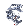
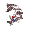
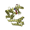
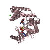
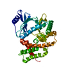
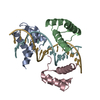
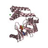
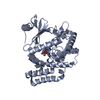
| Title | Aminoglycoside Phosphotransferase (2'')-Ia in complex with GMPPNP, Magnesium, and Lividomycin moieties | ||||||
 Components Components | Bifunctional AAC/APH | ||||||
 Keywords Keywords | TRANSFERASE/Antibiotic / Kinase / Antibiotic / Aminoglycoside / Resistance / Transferase / TRANSFERASE-Antibiotic complex | ||||||
| Function / homology |  Function and homology information Function and homology informationaminoglycoside 2''-phosphotransferase / aminoglycoside phosphotransferase activity / : / Transferases; Acyltransferases; Transferring groups other than aminoacyl groups / response to antibiotic / ATP binding / cytoplasm Similarity search - Function | ||||||
| Biological species |   Staphylococcus aureus (bacteria) Staphylococcus aureus (bacteria) | ||||||
| Method |  X-RAY DIFFRACTION / SYNCHROTRON / FOURIER SYNTHESIS / Resolution: 2.4 Å X-RAY DIFFRACTION / SYNCHROTRON / FOURIER SYNTHESIS / Resolution: 2.4 Å | ||||||
 Authors Authors | Caldwell, S.J. / Berghuis, A.M. | ||||||
| Funding support |  Canada, 1items Canada, 1items
| ||||||
 Citation Citation | Journal: Antimicrob. Agents Chemother. / Year: 2018 Title: Plasticity of Aminoglycoside Binding to Antibiotic Kinase APH(2′′)-Ia. Authors: Caldwell, S.J. / Berghuis, A.M. | ||||||
| History |
|
- Structure visualization
| Structure viewer | Molecule: MolmilJmol/JSmol |
|---|
- Downloads & links
-Download
| PDBx/mmCIF format | 6cey.cif.gz | 521.1 KB | Display | PDBx/mmCIF format |
|---|---|---|---|---|
| PDB format | pdb6cey.ent.gz | 428.3 KB | Display | PDB format |
| PDBx/mmJSON format | 6cey.json.gz | Tree view | PDBx/mmJSON format | |
| Others |  Other downloads Other downloads |
-Validation report
| Arichive directory | https://data.pdbj.org/pub/pdb/validation_reports/ce/6ceyftp://data.pdbj.org/pub/pdb/validation_reports/ce/6cey | HTTPS FTP |
|---|
-Related structure data
| Related structure data |  6c5uC 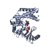 6cavC  6cgdC 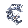 6cggC  6ch4C  5iqaS S: Starting model for refinement C: citing same article ( |
|---|---|
| Similar structure data |


-Links
 PDBj
PDBj


- Assembly
| Deposited unit | 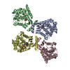
| ||||||||||||||||||||||||||||||||||||||||||||||||||||||||||||||||||||||||||||||||||||||||||||||||||||||||||||||||||||||||||||||||||||||||||||||||||||||||||||||||||||||||||||||||
|---|---|---|---|---|---|---|---|---|---|---|---|---|---|---|---|---|---|---|---|---|---|---|---|---|---|---|---|---|---|---|---|---|---|---|---|---|---|---|---|---|---|---|---|---|---|---|---|---|---|---|---|---|---|---|---|---|---|---|---|---|---|---|---|---|---|---|---|---|---|---|---|---|---|---|---|---|---|---|---|---|---|---|---|---|---|---|---|---|---|---|---|---|---|---|---|---|---|---|---|---|---|---|---|---|---|---|---|---|---|---|---|---|---|---|---|---|---|---|---|---|---|---|---|---|---|---|---|---|---|---|---|---|---|---|---|---|---|---|---|---|---|---|---|---|---|---|---|---|---|---|---|---|---|---|---|---|---|---|---|---|---|---|---|---|---|---|---|---|---|---|---|---|---|---|---|---|---|
| 1 | 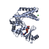
| ||||||||||||||||||||||||||||||||||||||||||||||||||||||||||||||||||||||||||||||||||||||||||||||||||||||||||||||||||||||||||||||||||||||||||||||||||||||||||||||||||||||||||||||||
| 2 | 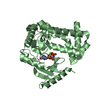
| ||||||||||||||||||||||||||||||||||||||||||||||||||||||||||||||||||||||||||||||||||||||||||||||||||||||||||||||||||||||||||||||||||||||||||||||||||||||||||||||||||||||||||||||||
| 3 | 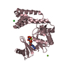
| ||||||||||||||||||||||||||||||||||||||||||||||||||||||||||||||||||||||||||||||||||||||||||||||||||||||||||||||||||||||||||||||||||||||||||||||||||||||||||||||||||||||||||||||||
| 4 | 
| ||||||||||||||||||||||||||||||||||||||||||||||||||||||||||||||||||||||||||||||||||||||||||||||||||||||||||||||||||||||||||||||||||||||||||||||||||||||||||||||||||||||||||||||||
| Unit cell |
| ||||||||||||||||||||||||||||||||||||||||||||||||||||||||||||||||||||||||||||||||||||||||||||||||||||||||||||||||||||||||||||||||||||||||||||||||||||||||||||||||||||||||||||||||
| Noncrystallographic symmetry (NCS) | NCS domain:
NCS domain segments: Component-ID: _ / Refine code: _
NCS ensembles :
|
-Components
-Protein , 1 types, 4 molecules ABCD
| #1: Protein | Mass: 35948.199 Da / Num. of mol.: 4 Source method: isolated from a genetically manipulated source Source: (gene. exp.) Staphylococcus aureus (bacteria) / Gene: aacA-aphD, R015, VRA0030 / Plasmid: pET-22b-APH(2'')-Ia / Production host: References: UniProt: P0A0C1, Transferases; Acyltransferases; Transferring groups other than aminoacyl groups, aminoglycoside 2''-phosphotransferase |
|---|
-Non-polymers , 5 types, 558 molecules 


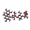

| #2: Chemical | ChemComp-GNP /  Mass: 522.196 Da / Num. of mol.: 4 / Source method: obtained synthetically / Formula: C10H17N6O13P3 Mass: 522.196 Da / Num. of mol.: 4 / Source method: obtained synthetically / Formula: C10H17N6O13P3Comment: GppNHp, GMPPNP, energy-carrying molecule analogue*YM #3: Chemical | ChemComp-MG /  Mass: 24.305 Da / Num. of mol.: 9 / Source method: obtained synthetically / Formula: Mg Mass: 24.305 Da / Num. of mol.: 9 / Source method: obtained synthetically / Formula: Mg#4: Chemical |  Mass: 35.453 Da / Num. of mol.: 3 / Source method: obtained synthetically / Formula: Cl Mass: 35.453 Da / Num. of mol.: 3 / Source method: obtained synthetically / Formula: Cl#5: Chemical | ChemComp-LIV / ( |  Mass: 761.770 Da / Num. of mol.: 1 / Source method: obtained synthetically / Formula: C29H55N5O18 Mass: 761.770 Da / Num. of mol.: 1 / Source method: obtained synthetically / Formula: C29H55N5O18#6: Water | ChemComp-HOH / | Mass: 18.015 Da / Num. of mol.: 541 / Source method: isolated from a natural source / Formula: H2O |
|---|
-Experimental details
-Experiment
| Experiment | Method: X-RAY DIFFRACTION / Number of used crystals: 1 |
|---|
- Sample preparation
| Crystal | Density Matthews: 2.87 Å3/Da / Density % sol: 56.91 % |
|---|---|
| Crystal grow | Temperature: 296 K / Method: vapor diffusion, hanging drop / pH: 7.5 Details: 80-120mM MgCl2, 8% glycerol, 10% PEG 3350, 100mM HEPES pH 7.5 |
-Data collection
| Diffraction | Mean temperature: 100 K |
|---|---|
| Diffraction source | Source: SYNCHROTRON / Site: CLSI / Beamline: 08ID-1 / Wavelength: 0.9795 Å |
| Detector | Type: RAYONIX MX-300 / Detector: CCD / Date: Nov 26, 2013 |
| Radiation | Monochromator: ACCEL/BRUKER double crystal monochromator (DCM), featuring indirectly cryo-cooled first crystal and sagittally focusing second crystal Protocol: SINGLE WAVELENGTH / Monochromatic (M) / Laue (L): M / Scattering type: x-ray |
| Radiation wavelength | Wavelength: 0.9795 Å / Relative weight: 1 |
| Reflection | Resolution: 2.4→90.73 Å / Num. obs: 63302 / % possible obs: 100 % / Redundancy: 4.2 % / Biso Wilson estimate: 50.686 Å2 / CC1/2: 0.988 / Rmerge(I) obs: 0.11 / Rpim(I) all: 0.084 / Rrim(I) all: 0.127 / Net I/σ(I): 10 |
| Reflection shell | Resolution: 2.4→2.46 Å / Redundancy: 4.2 % / Rmerge(I) obs: 1.345 / Mean I/σ(I) obs: 1.8 / Num. unique obs: 4441 / CC1/2: 0.287 / Rpim(I) all: 1.011 / Rrim(I) all: 1.538 / % possible all: 100 |
- Processing
| Software |
| ||||||||||||||||||||||||||||||||||||||||||||||||||||||||||||||||||||||||||||||||||||||||||||||||||||||||||||||||||||||||||||||||||||||||||||||||||||||||||||||||||||||||||||||||||||||
|---|---|---|---|---|---|---|---|---|---|---|---|---|---|---|---|---|---|---|---|---|---|---|---|---|---|---|---|---|---|---|---|---|---|---|---|---|---|---|---|---|---|---|---|---|---|---|---|---|---|---|---|---|---|---|---|---|---|---|---|---|---|---|---|---|---|---|---|---|---|---|---|---|---|---|---|---|---|---|---|---|---|---|---|---|---|---|---|---|---|---|---|---|---|---|---|---|---|---|---|---|---|---|---|---|---|---|---|---|---|---|---|---|---|---|---|---|---|---|---|---|---|---|---|---|---|---|---|---|---|---|---|---|---|---|---|---|---|---|---|---|---|---|---|---|---|---|---|---|---|---|---|---|---|---|---|---|---|---|---|---|---|---|---|---|---|---|---|---|---|---|---|---|---|---|---|---|---|---|---|---|---|---|---|
| Refinement | Method to determine structure: FOURIER SYNTHESIS Starting model: PDB 5IQA Resolution: 2.4→90.73 Å / Cor.coef. Fo:Fc: 0.966 / Cor.coef. Fo:Fc free: 0.947 / SU B: 16.142 / SU ML: 0.18 / Cross valid method: THROUGHOUT / ESU R: 0.312 / ESU R Free: 0.219 / Stereochemistry target values: MAXIMUM LIKELIHOOD Details: Author states that the ligand modelled is their best interpretation of the observed electron density with the tools available at deposition, but remains subject to some ambiguity in absolute ...Details: Author states that the ligand modelled is their best interpretation of the observed electron density with the tools available at deposition, but remains subject to some ambiguity in absolute configuration. The difference density was observed following the soaking of lividomycin to antibiotic-free crystals. Observed electron density likely reflects the superimposition of multiple weak binding modes, which would explain to low-level off-target activity of the enzyme as reported in studies of enzymatic regiospecificity.
| ||||||||||||||||||||||||||||||||||||||||||||||||||||||||||||||||||||||||||||||||||||||||||||||||||||||||||||||||||||||||||||||||||||||||||||||||||||||||||||||||||||||||||||||||||||||
| Solvent computation | Ion probe radii: 0.8 Å / Shrinkage radii: 0.8 Å / VDW probe radii: 1.2 Å / Solvent model: MASK | ||||||||||||||||||||||||||||||||||||||||||||||||||||||||||||||||||||||||||||||||||||||||||||||||||||||||||||||||||||||||||||||||||||||||||||||||||||||||||||||||||||||||||||||||||||||
| Displacement parameters | Biso mean: 59.493 Å2
| ||||||||||||||||||||||||||||||||||||||||||||||||||||||||||||||||||||||||||||||||||||||||||||||||||||||||||||||||||||||||||||||||||||||||||||||||||||||||||||||||||||||||||||||||||||||
| Refinement step | Cycle: 1 / Resolution: 2.4→90.73 Å
| ||||||||||||||||||||||||||||||||||||||||||||||||||||||||||||||||||||||||||||||||||||||||||||||||||||||||||||||||||||||||||||||||||||||||||||||||||||||||||||||||||||||||||||||||||||||
| Refine LS restraints |
|