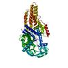
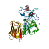
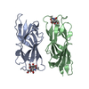
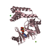
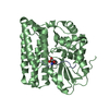
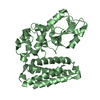
- PDB-3vgo: Crystal structure of the N-terminal fragment of Cbl-b -
+
Open data
ID or keywords:
Loading...
-
Basic information
Entry
Database: PDB / ID: 3vgo
Title
Crystal structure of the N-terminal fragment of Cbl-b
Components
E3 ubiquitin-protein ligase CBL-B
Keywords
LIGASE / MEROHEDRAL TWINNING / E3 ligase
Function / homology
Function and homology information
regulation of platelet-derived growth factor receptor-alpha signaling pathway / T cell anergy / regulation protein catabolic process at postsynapse / positive regulation of T cell anergy / CD4-positive, alpha-beta T cell proliferation / negative regulation of CD4-positive, alpha-beta T cell proliferation / NLS-bearing protein import into nucleus / regulation of postsynaptic neurotransmitter receptor internalization / negative regulation of T cell activation / negative regulation of epidermal growth factor receptor signaling pathway ...regulation of platelet-derived growth factor receptor-alpha signaling pathway / T cell anergy / regulation protein catabolic process at postsynapse / positive regulation of T cell anergy / CD4-positive, alpha-beta T cell proliferation / negative regulation of CD4-positive, alpha-beta T cell proliferation / NLS-bearing protein import into nucleus / regulation of postsynaptic neurotransmitter receptor internalization / negative regulation of T cell activation / negative regulation of epidermal growth factor receptor signaling pathway / negative regulation of T cell receptor signaling pathway / protein K63-linked ubiquitination / phosphotyrosine residue binding / positive regulation of protein ubiquitination / protein catabolic process / receptor tyrosine kinase binding / RING-type E3 ubiquitin transferase / positive regulation of protein catabolic process / ubiquitin protein ligase activity / T cell receptor signaling pathway / Antigen processing: Ubiquitination & Proteasome degradation / protein stabilization / intracellular signal transduction / postsynapse / immune response / membrane raft / calcium ion binding / glutamatergic synapse / signal transduction / zinc ion binding / nucleoplasm / plasma membrane / cytosol Similarity search - Function
E3 ubiquitin-protein ligase CBL-B, RING finger, HC subclass / Adaptor protein Cbl, N-terminal domain / Adaptor protein Cbl, N-terminal helical / Adaptor protein Cbl, EF hand-like / Adaptor protein Cbl, SH2-like domain / Adaptor protein Cbl, PTB domain / Adaptor protein Cbl / CBL proto-oncogene N-terminal domain 1 / CBL proto-oncogene N-terminus, EF hand-like domain / CBL proto-oncogene N-terminus, SH2-like domain ...E3 ubiquitin-protein ligase CBL-B, RING finger, HC subclass / Adaptor protein Cbl, N-terminal domain / Adaptor protein Cbl, N-terminal helical / Adaptor protein Cbl, EF hand-like / Adaptor protein Cbl, SH2-like domain / Adaptor protein Cbl, PTB domain / Adaptor protein Cbl / CBL proto-oncogene N-terminal domain 1 / CBL proto-oncogene N-terminus, EF hand-like domain / CBL proto-oncogene N-terminus, SH2-like domain / Cbl-type phosphotyrosine-binding (Cbl-PTB) domain profile. / Prokaryotic RING finger family 4 / Adaptor protein Cbl, N-terminal domain superfamily / Transcription Elongation Factor S-II; Chain A / Ubiquitin associated domain / Ubiquitin-associated domain / Ubiquitin-associated domain (UBA) profile. / SH2 domain / Zinc finger, C3HC4 RING-type / SHC Adaptor Protein / Zinc finger, C3HC4 type (RING finger) / EF-hand / Recoverin; domain 1 / Zinc finger, RING-type, conserved site / Zinc finger RING-type signature. / Ring finger / SH2 domain superfamily / Zinc finger RING-type profile. / Zinc finger, RING-type / EF-hand domain pair / Zinc finger, RING/FYVE/PHD-type / Up-down Bundle / 2-Layer Sandwich / Orthogonal Bundle / Mainly Alpha / Alpha Beta Similarity search - Domain/homology
Resolution: 3.1→42.82 Å / Data cutoff high absF: 57063.23 / Data cutoff low absF: 0 / Isotropic thermal model: GROUP / Cross valid method: THROUGHOUT / σ(F): 0 / Stereochemistry target values: Engh & Huber Details: BULK SOLVENT MODEL USED. CNS script refine_twin.inp was used for refinement.
Rfactor
Num. reflection
% reflection
Selection details
Rfree
0.328
1909
9.4 %
CNS SCRIPT MAKE_CV_TWIN.INP, ENSURES THAT TWIN-RELATED PAIRS OF REFLECTIONS ARE EITHER BOTH IN TEST OR BOTH IN WORK SET
In the structure databanks used in Yorodumi, some data are registered as the other names, "COVID-19 virus" and "2019-nCoV". Here are the details of the virus and the list of structure data.
Jan 31, 2019. EMDB accession codes are about to change! (news from PDBe EMDB page)
EMDB accession codes are about to change! (news from PDBe EMDB page)
The allocation of 4 digits for EMDB accession codes will soon come to an end. Whilst these codes will remain in use, new EMDB accession codes will include an additional digit and will expand incrementally as the available range of codes is exhausted. The current 4-digit format prefixed with “EMD-” (i.e. EMD-XXXX) will advance to a 5-digit format (i.e. EMD-XXXXX), and so on. It is currently estimated that the 4-digit codes will be depleted around Spring 2019, at which point the 5-digit format will come into force.
The EM Navigator/Yorodumi systems omit the EMD- prefix.
Related info.:Q: What is EMD? / ID/Accession-code notation in Yorodumi/EM Navigator
Yorodumi is a browser for structure data from EMDB, PDB, SASBDB, etc.
This page is also the successor to EM Navigator detail page, and also detail information page/front-end page for Omokage search.
The word "yorodu" (or yorozu) is an old Japanese word meaning "ten thousand". "mi" (miru) is to see.
Related info.:EMDB / PDB / SASBDB / Comparison of 3 databanks / Yorodumi Search / Aug 31, 2016. New EM Navigator & Yorodumi / Yorodumi Papers / Jmol/JSmol / Function and homology information / Changes in new EM Navigator and Yorodumi
 Movie
Movie Controller


 Open data
 Basic information
 Components
 Keywords
 Function and homology information
 Homo sapiens (human)
 X-RAY DIFFRACTION /
  Authors
 Citation
 Structure visualization
 Downloads & links
 Other downloads


 PDBj
PDBj

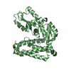
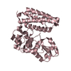
 Assembly
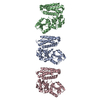


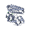
 Sample preparation
 Processing