 Movie
Movie Controller

[English] 日本語
 Yorodumi
- PDB-1fch: CRYSTAL STRUCTURE OF THE PTS1 COMPLEXED TO THE TPR REGION OF HUMA... -
+ Open data

- Basic information
| Entry | Database: PDB / ID: 1fch | ||||||
|---|---|---|---|---|---|---|---|
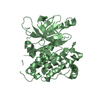
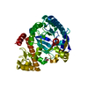
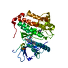
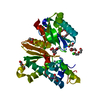
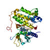
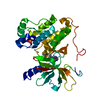
| Title | CRYSTAL STRUCTURE OF THE PTS1 COMPLEXED TO THE TPR REGION OF HUMAN PEX5 | ||||||
 Components Components |
| ||||||
 Keywords Keywords | SIGNALING PROTEIN / PROTEIN-PEPTIDE COMPLEX / TETRATRICOPEPTIDE REPEAT / TPR / HELICAL REPEAT | ||||||
| Function / homology |  Function and homology information Function and homology informationprotein import into peroxisome matrix, substrate release / protein import into peroxisome matrix, translocation / peroxisome membrane targeting sequence binding / protein import into peroxisome membrane / protein targeting to peroxisome / peroxisome matrix targeting signal-1 binding / peroxisome signal sequence receptor activity / protein import into peroxisome matrix, receptor recycling / protein import into peroxisome matrix / protein import into peroxisome matrix, docking ...protein import into peroxisome matrix, substrate release / protein import into peroxisome matrix, translocation / peroxisome membrane targeting sequence binding / protein import into peroxisome membrane / protein targeting to peroxisome / peroxisome matrix targeting signal-1 binding / peroxisome signal sequence receptor activity / protein import into peroxisome matrix, receptor recycling / protein import into peroxisome matrix / protein import into peroxisome matrix, docking / protein carrier activity / very long-chain fatty acid metabolic process / cerebral cortex neuron differentiation / cell development / pexophagy / endoplasmic reticulum organization / positive regulation of multicellular organism growth / peroxisomal membrane / mitochondrial membrane organization / neuromuscular process / fatty acid beta-oxidation / cerebral cortex cell migration / peroxisomal matrix / negative regulation of protein-containing complex assembly / Pexophagy / cellular response to reactive oxygen species / protein tetramerization / Peroxisomal protein import / neuron migration / small GTPase binding / peroxisome / E3 ubiquitin ligases ubiquitinate target proteins / enzyme binding / Golgi apparatus / protein-containing complex / mitochondrion / membrane / cytoplasm / cytosol Similarity search - Function | ||||||
| Biological species |  Homo sapiens (human) Homo sapiens (human) | ||||||
| Method |  X-RAY DIFFRACTION / Resolution: 2.2 Å X-RAY DIFFRACTION / Resolution: 2.2 Å | ||||||
 Authors Authors | Gatto Jr., G.J. / Geisbrecht, B.V. / Gould, S.J. / Berg, J.M. | ||||||
 Citation Citation | Journal: Nat.Struct.Biol. / Year: 2000 Title: Peroxisomal targeting signal-1 recognition by the TPR domains of human PEX5. Authors: Gatto Jr., G.J. / Geisbrecht, B.V. / Gould, S.J. / Berg, J.M. | ||||||
| History |
|
- Structure visualization
| Structure viewer | Molecule: MolmilJmol/JSmol |
|---|
- Downloads & links
-Download
| PDBx/mmCIF format | 1fch.cif.gz | 132.6 KB | Display | PDBx/mmCIF format |
|---|---|---|---|---|
| PDB format | pdb1fch.ent.gz | 103.7 KB | Display | PDB format |
| PDBx/mmJSON format | 1fch.json.gz | Tree view | PDBx/mmJSON format | |
| Others |  Other downloads Other downloads |
-Validation report
| Arichive directory | https://data.pdbj.org/pub/pdb/validation_reports/fc/1fchftp://data.pdbj.org/pub/pdb/validation_reports/fc/1fch | HTTPS FTP |
|---|
-Related structure data
| Similar structure data |
|---|


-Links
 PDBj
PDBj


- Assembly
| Deposited unit | 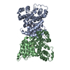
| ||||||||
|---|---|---|---|---|---|---|---|---|---|
| 1 | 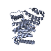
| ||||||||
| 2 | 
| ||||||||
| 3 | 
| ||||||||
| 4 |
| ||||||||
| 5 | 
| ||||||||
| Unit cell |
|
-Components
| #1: Protein | Mass: 40989.910 Da / Num. of mol.: 2 / Fragment: C-TERMINAL TPR REGION Source method: isolated from a genetically manipulated source Source: (gene. exp.) Homo sapiens (human) / Plasmid: PT7 / Production host:  #2: Protein/peptide | Mass: 638.733 Da / Num. of mol.: 2 / Source method: obtained synthetically / Details: THE PEPTIDE WAS CHEMICALLY SYNTHESIZED #3: Water | ChemComp-HOH / |  Mass: 18.015 Da / Num. of mol.: 241 / Source method: isolated from a natural source / Formula: H2O Mass: 18.015 Da / Num. of mol.: 241 / Source method: isolated from a natural source / Formula: H2O |
|---|
-Experimental details
-Experiment
| Experiment | Method: X-RAY DIFFRACTION / Number of used crystals: 1 |
|---|
- Sample preparation
| Crystal | Density Matthews: 2.29 Å3/Da / Density % sol: 46.29 % | ||||||||||||||||||||||||||||||||||||||||||
|---|---|---|---|---|---|---|---|---|---|---|---|---|---|---|---|---|---|---|---|---|---|---|---|---|---|---|---|---|---|---|---|---|---|---|---|---|---|---|---|---|---|---|---|
| Crystal grow | Temperature: 293 K / Method: vapor diffusion, hanging drop / pH: 7.5 Details: Sodium HEPES, Sodium Citrate, pH 7.5, VAPOR DIFFUSION, HANGING DROP, temperature 293K | ||||||||||||||||||||||||||||||||||||||||||
| Crystal grow | *PLUS Details: peptide solution was diluted 1:1 with reservoir | ||||||||||||||||||||||||||||||||||||||||||
| Components of the solutions | *PLUS
|
-Data collection
| Diffraction | Mean temperature: 93 K |
|---|---|
| Diffraction source | Source: ROTATING ANODE / Type: RIGAKU RU200 / Wavelength: 1.5418 |
| Detector | Type: RIGAKU RAXIS II / Detector: IMAGE PLATE / Date: May 9, 2000 |
| Radiation | Protocol: SINGLE WAVELENGTH / Monochromatic (M) / Laue (L): M / Scattering type: x-ray |
| Radiation wavelength | Wavelength: 1.5418 Å / Relative weight: 1 |
| Reflection | Resolution: 2.2→30 Å / Num. all: 38298 / Num. obs: 38298 / % possible obs: 99.8 % / Redundancy: 3.6 % / Biso Wilson estimate: 21.2 Å2 / Rmerge(I) obs: 0.055 / Net I/σ(I): 21.8 |
| Reflection shell | Resolution: 2.2→2.24 Å / Redundancy: 3.1 % / Rmerge(I) obs: 0.227 / Num. unique all: 1916 / % possible all: 98.8 |
| Reflection shell | *PLUS % possible obs: 98.8 % / Mean I/σ(I) obs: 5.9 |
- Processing
| Software |
| ||||||||||||||||||||||||||||||||||||
|---|---|---|---|---|---|---|---|---|---|---|---|---|---|---|---|---|---|---|---|---|---|---|---|---|---|---|---|---|---|---|---|---|---|---|---|---|---|
| Refinement | Resolution: 2.2→29.63 Å / Rfactor Rfree error: 0.004 / Data cutoff high absF: 2170352.49 / Data cutoff low absF: 0 / Isotropic thermal model: RESTRAINED / Cross valid method: THROUGHOUT / σ(F): 0
| ||||||||||||||||||||||||||||||||||||
| Solvent computation | Solvent model: FLAT MODEL / Bsol: 33.56 Å2 / ksol: 0.351 e/Å3 | ||||||||||||||||||||||||||||||||||||
| Displacement parameters | Biso mean: 30.4 Å2
| ||||||||||||||||||||||||||||||||||||
| Refine analyze |
| ||||||||||||||||||||||||||||||||||||
| Refinement step | Cycle: LAST / Resolution: 2.2→29.63 Å
| ||||||||||||||||||||||||||||||||||||
| Refine LS restraints |
| ||||||||||||||||||||||||||||||||||||
| LS refinement shell | Resolution: 2.2→2.34 Å / Rfactor Rfree error: 0.01 / Total num. of bins used: 6
| ||||||||||||||||||||||||||||||||||||
| Xplor file |
| ||||||||||||||||||||||||||||||||||||
| Software | *PLUS Name: CNS / Version: 1 / Classification: refinement | ||||||||||||||||||||||||||||||||||||
| Refine LS restraints | *PLUS
|
