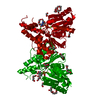
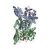
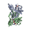
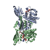
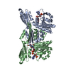
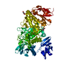
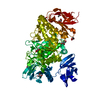
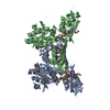
[English] 日本語
Yorodumi
- PDB-2yhw: High-resolution crystal structures of N-Acetylmannosamine kinase:... -

+
Open data


ID or keywords:

Loading...

-
Basic information

Entry
Database: PDB / ID: 2yhw
TitleHigh-resolution crystal structures of N-Acetylmannosamine kinase: Insights about substrate specificity, activity and inhibitor modelling.
ComponentsBIFUNCTIONAL UDP-N-ACETYLGLUCOSAMINE 2-EPIMERASE/N-ACETYLMANNOSAMINE KINASE
KeywordsTRANSFERASE / SIALIC ACID / MANNAC / ROK FAMILY
Function / homology
Function and homology information


Defective GNE causes sialuria, NK and IBM2 / UDP-N-acetylglucosamine 2-epimerase (hydrolysing) / N-acetylglucosamine biosynthetic process / N-acylmannosamine kinase / N-acylmannosamine kinase activity / N-acetylneuraminate biosynthetic process / hexokinase activity / UDP-N-acetylglucosamine 2-epimerase activity / UDP-N-acetylglucosamine metabolic process / Sialic acid metabolism ...Defective GNE causes sialuria, NK and IBM2 / UDP-N-acetylglucosamine 2-epimerase (hydrolysing) / N-acetylglucosamine biosynthetic process / N-acylmannosamine kinase / N-acylmannosamine kinase activity / N-acetylneuraminate biosynthetic process / hexokinase activity / UDP-N-acetylglucosamine 2-epimerase activity / UDP-N-acetylglucosamine metabolic process / Sialic acid metabolism / hydrolase activity, hydrolyzing O-glycosyl compounds / ATP binding / metal ion binding / cytosol
Similarity search - Function
UDP-N-acetylglucosamine 2-epimerase,UDP-hydrolysing / UDP-N-acetylglucosamine 2-epimerase domain / UDP-N-acetylglucosamine 2-epimerase / ROK family / ROK family / ATPase, nucleotide binding domain / ATPase, nucleotide binding domain / Nucleotidyltransferase; domain 5 / 2-Layer Sandwich / Alpha Beta
Similarity search - Domain/homology
ACETATE ION / 2-acetamido-2-deoxy-alpha-D-mannopyranose / TRIETHYLENE GLYCOL / Bifunctional UDP-N-acetylglucosamine 2-epimerase/N-acetylmannosamine kinase
Similarity search - Component
Biological speciesHOMO SAPIENS (human)
MethodX-RAY DIFFRACTION / SYNCHROTRON / OTHER / Resolution: 1.64 Å
AuthorsMartinez, J. / Nguyen, L.D. / Tauberger, E. / Hinderlich, S. / Zimmer, R. / Tauberger, E. / Reutter, W. / Saenger, W. / Fan, H. / Moniot, S.
CitationJournal: J.Biol.Chem. / Year: 2012
Title: Crystal Structures of N-Acetylmannosamine Kinase Provide Insights Into Enzyme Specificity and Inhibition
Authors: Martinez, J. / Nguyen, L.D. / Tauberger, E. / Hinderlich, S. / Reutter, W. / Fan, H. / Saenger, W. / Moniot, S.
History
DepositionMay 8, 2011Deposition site: PDBE / Processing site: PDBE
Revision 1.0Feb 29, 2012Provider: repository / Type: Initial release
Revision 1.1May 2, 2012Group: Other
Revision 1.2Jul 29, 2020Group: Data collection / Derived calculations ...Data collection / Derived calculations / Other / Structure summary
Category: chem_comp / entity ...chem_comp / entity / pdbx_chem_comp_identifier / pdbx_database_status / pdbx_entity_nonpoly / pdbx_struct_conn_angle / struct_conn / struct_site / struct_site_gen
Item: _chem_comp.name / _chem_comp.type ..._chem_comp.name / _chem_comp.type / _entity.pdbx_description / _pdbx_database_status.status_code_sf / _pdbx_entity_nonpoly.name / _pdbx_struct_conn_angle.ptnr1_auth_comp_id / _pdbx_struct_conn_angle.ptnr1_auth_seq_id / _pdbx_struct_conn_angle.ptnr1_label_asym_id / _pdbx_struct_conn_angle.ptnr1_label_atom_id / _pdbx_struct_conn_angle.ptnr1_label_comp_id / _pdbx_struct_conn_angle.ptnr1_label_seq_id / _pdbx_struct_conn_angle.ptnr2_auth_comp_id / _pdbx_struct_conn_angle.ptnr2_auth_seq_id / _pdbx_struct_conn_angle.ptnr2_label_alt_id / _pdbx_struct_conn_angle.ptnr2_label_asym_id / _pdbx_struct_conn_angle.ptnr2_label_atom_id / _pdbx_struct_conn_angle.ptnr2_label_comp_id / _pdbx_struct_conn_angle.ptnr3_auth_comp_id / _pdbx_struct_conn_angle.ptnr3_auth_seq_id / _pdbx_struct_conn_angle.ptnr3_label_asym_id / _pdbx_struct_conn_angle.ptnr3_label_atom_id / _pdbx_struct_conn_angle.ptnr3_label_comp_id / _pdbx_struct_conn_angle.ptnr3_label_seq_id / _pdbx_struct_conn_angle.value / _struct_conn.pdbx_dist_value / _struct_conn.pdbx_leaving_atom_flag / _struct_conn.pdbx_ptnr1_label_alt_id / _struct_conn.pdbx_ptnr2_label_alt_id / _struct_conn.ptnr1_auth_comp_id / _struct_conn.ptnr1_auth_seq_id / _struct_conn.ptnr1_label_asym_id / _struct_conn.ptnr1_label_atom_id / _struct_conn.ptnr1_label_comp_id / _struct_conn.ptnr1_label_seq_id / _struct_conn.ptnr2_auth_comp_id / _struct_conn.ptnr2_auth_seq_id / _struct_conn.ptnr2_label_asym_id / _struct_conn.ptnr2_label_atom_id / _struct_conn.ptnr2_label_comp_id / _struct_conn.ptnr2_label_seq_id
Description: Carbohydrate remediation / Provider: repository / Type: Remediation
Revision 2.0Nov 15, 2023Group: Atomic model / Data collection ...Atomic model / Data collection / Database references / Derived calculations / Structure summary
Category: atom_site / atom_site_anisotrop ...atom_site / atom_site_anisotrop / chem_comp / chem_comp_atom / chem_comp_bond / database_2 / struct_conn
Item: _atom_site.auth_atom_id / _atom_site.label_atom_id ..._atom_site.auth_atom_id / _atom_site.label_atom_id / _atom_site_anisotrop.pdbx_auth_atom_id / _atom_site_anisotrop.pdbx_label_atom_id / _chem_comp.pdbx_synonyms / _database_2.pdbx_DOI / _database_2.pdbx_database_accession / _struct_conn.pdbx_leaving_atom_flag / _struct_conn.ptnr2_label_atom_id

-
Structure visualization

Structure viewerMolecule:
MolmilJmol/JSmol

Downloads & links

-
Assembly

Deposited unit
A: BIFUNCTIONAL UDP-N-ACETYLGLUCOSAMINE 2-EPIMERASE/N-ACETYLMANNOSAMINE KINASE
hetero molecules


Theoretical massNumber of molelcules
Total (without water)38,00413
Polymers36,6201
Non-polymers1,38412
Water4,594255
1
A: BIFUNCTIONAL UDP-N-ACETYLGLUCOSAMINE 2-EPIMERASE/N-ACETYLMANNOSAMINE KINASE
hetero molecules

A: BIFUNCTIONAL UDP-N-ACETYLGLUCOSAMINE 2-EPIMERASE/N-ACETYLMANNOSAMINE KINASE
hetero molecules


Theoretical massNumber of molelcules
Total (without water)76,00926
Polymers73,2402
Non-polymers2,76924
Water362
TypeNameSymmetry operationNumber
identity operation1_555x,y,z1
crystal symmetry operation8_555-y,-x,-z+1/21
Buried area8040 Å2
ΔGint-31.3 kcal/mol
Surface area25110 Å2
MethodPISA
Unit cell
Length a, b, c (Å)90.730, 90.730, 100.780
Angle α, β, γ (deg.)90.00, 90.00, 90.00
Int Tables number92
Space group name H-MP41212

-
Components

-
Protein / Sugars , 2 types, 2 molecules A

#1: Protein BIFUNCTIONAL UDP-N-ACETYLGLUCOSAMINE 2-EPIMERASE/N-ACETYLMANNOSAMINE KINASE / N-ACETYL MANNOSAMINE KINASE / MANAC KINASE / UDP-GLCNAC-2-EPIMERASE/MANAC KINASE


Mass: 36620.016 Da / Num. of mol.: 1
Fragment: N-ACETYLMANNOSAMINE KINASE DOMAIN, RESIDUES 406-720
Source method: isolated from a genetically manipulated source
Source: (gene. exp.) HOMO SAPIENS (human) / Plasmid: PET28A-MNK / Production host: ESCHERICHIA COLI (E. coli) / Strain (production host): BL21(DE3) / Variant (production host): RIL / References: UniProt: Q9Y223, N-acylmannosamine kinase
#2: Sugar ChemComp-BM3 / 2-acetamido-2-deoxy-alpha-D-mannopyranose / N-acetyl-alpha-D-mannosamine / 2-acetamido-2-deoxy-alpha-D-mannose / 2-acetamido-2-deoxy-D-mannose / 2-acetamido-2-deoxy-mannose / 2-(ACETYLAMINO)-2-DEOXY-ALPHA-D-MANNOPYRANOSE


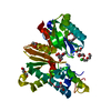
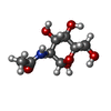
Type: D-saccharide, alpha linking / Mass: 221.208 Da / Num. of mol.: 1 / Source method: obtained synthetically / Formula: C8H15NO6
IdentifierTypeProgram
DManpNAcaCONDENSED IUPAC CARBOHYDRATE SYMBOLGMML 1.0
N-acetyl-a-D-mannopyranosamineCOMMON NAMEGMML 1.0
a-D-ManpNAcIUPAC CARBOHYDRATE SYMBOLPDB-CARE 1.0
ManNAcSNFG CARBOHYDRATE SYMBOLGMML 1.0

-
Non-polymers , 8 types, 266 molecules

#3: Chemical ChemComp-ZN / ZINC ION


Mass: 65.409 Da / Num. of mol.: 1 / Source method: obtained synthetically / Formula: Zn
#4: Chemical ChemComp-2PE / NONAETHYLENE GLYCOL


Mass: 414.488 Da / Num. of mol.: 1 / Source method: obtained synthetically / Formula: C18H38O10 / Comment: precipitant*YM
#5: Chemical
ChemComp-EDO / 1,2-ETHANEDIOL / ETHYLENE GLYCOL


Mass: 62.068 Da / Num. of mol.: 4 / Source method: obtained synthetically / Formula: C2H6O2
#6: Chemical ChemComp-CA / CALCIUM ION


Mass: 40.078 Da / Num. of mol.: 1 / Source method: obtained synthetically / Formula: Ca
#7: Chemical ChemComp-PGE / TRIETHYLENE GLYCOL


Mass: 150.173 Da / Num. of mol.: 2 / Source method: obtained synthetically / Formula: C6H14O4
#8: Chemical ChemComp-ACT / ACETATE ION


Mass: 59.044 Da / Num. of mol.: 1 / Source method: obtained synthetically / Formula: C2H3O2
#9: Chemical ChemComp-CL / CHLORIDE ION


Mass: 35.453 Da / Num. of mol.: 1 / Source method: obtained synthetically / Formula: Cl
#10: Water ChemComp-HOH / water


Mass: 18.015 Da / Num. of mol.: 255 / Source method: isolated from a natural source / Formula: H2O

-
Experimental details

-
Experiment

ExperimentMethod: X-RAY DIFFRACTION

-
Sample preparation

CrystalDensity Matthews: 2.83 Å3/Da / Density % sol: 56.57 % / Description: NONE
Crystal growDetails: 0.2 M CALCIUM ACETATE, 0.1 M SODIUM CACODYLATE PH 6.5 AND 40% PEG 300.

-
Data collection

DiffractionMean temperature: 100 K
Diffraction sourceSource: SYNCHROTRON / Site: BESSY / Beamline: 14.2 / Wavelength: 0.918
DetectorType: MARMOSAIC 225 mm CCD / Detector: CCD
RadiationProtocol: SINGLE WAVELENGTH / Monochromatic (M) / Laue (L): M / Scattering type: x-ray
Radiation wavelengthWavelength: 0.918 Å / Relative weight: 1
ReflectionResolution: 1.64→45.3 Å / Num. obs: 52083 / % possible obs: 100 % / Observed criterion σ(I): 4.37 / Redundancy: 12.9 % / Rmerge(I) obs: 0.06 / Net I/σ(I): 30.66

-
Processing

Software
NameVersionClassification
REFMAC5.5.0109refinement
XDSdata reduction
XSCALEdata scaling
RefinementMethod to determine structure: OTHER
Starting model: NONE

Resolution: 1.64→45.37 Å / Cor.coef. Fo:Fc: 0.97 / Cor.coef. Fo:Fc free: 0.965 / SU B: 2.27 / SU ML: 0.039 / Cross valid method: THROUGHOUT / ESU R: 0.067 / ESU R Free: 0.068 / Stereochemistry target values: MAXIMUM LIKELIHOOD / Details: HYDROGENS HAVE BEEN ADDED IN THE RIDING POSITIONS.
RfactorNum. reflection% reflectionSelection details
Rfree0.17019 2605 5 %RANDOM
Rwork0.1482 ---
obs0.14933 49478 100 %-
Solvent computationIon probe radii: 0.8 Å / Shrinkage radii: 0.8 Å / VDW probe radii: 1.4 Å / Solvent model: MASK
Displacement parametersBiso mean: 25.699 Å2
Baniso -1Baniso -2Baniso -3
1-0.39 Å20 Å20 Å2
2--0.39 Å20 Å2
3----0.79 Å2
Refinement stepCycle: LAST / Resolution: 1.64→45.37 Å
ProteinNucleic acidLigandSolventTotal
Num. atoms2271 0 71 255 2597
Refine LS restraints
Refine-IDTypeDev idealDev ideal targetNumber
X-RAY DIFFRACTIONr_bond_refined_d0.0260.0212583
X-RAY DIFFRACTIONr_bond_other_d0.0010.021678
X-RAY DIFFRACTIONr_angle_refined_deg2.0681.9823506
X-RAY DIFFRACTIONr_angle_other_deg1.18434160
X-RAY DIFFRACTIONr_dihedral_angle_1_deg6.6375349
X-RAY DIFFRACTIONr_dihedral_angle_2_deg37.2132596
X-RAY DIFFRACTIONr_dihedral_angle_3_deg14.31615432
X-RAY DIFFRACTIONr_dihedral_angle_4_deg19.7491512
X-RAY DIFFRACTIONr_chiral_restr0.1570.2418
X-RAY DIFFRACTIONr_gen_planes_refined0.010.022936
X-RAY DIFFRACTIONr_gen_planes_other0.0040.02470
X-RAY DIFFRACTIONr_nbd_refined
X-RAY DIFFRACTIONr_nbd_other
X-RAY DIFFRACTIONr_nbtor_refined
X-RAY DIFFRACTIONr_nbtor_other
X-RAY DIFFRACTIONr_xyhbond_nbd_refined
X-RAY DIFFRACTIONr_xyhbond_nbd_other
X-RAY DIFFRACTIONr_metal_ion_refined
X-RAY DIFFRACTIONr_metal_ion_other
X-RAY DIFFRACTIONr_symmetry_vdw_refined
X-RAY DIFFRACTIONr_symmetry_vdw_other
X-RAY DIFFRACTIONr_symmetry_hbond_refined
X-RAY DIFFRACTIONr_symmetry_hbond_other
X-RAY DIFFRACTIONr_symmetry_metal_ion_refined
X-RAY DIFFRACTIONr_symmetry_metal_ion_other
X-RAY DIFFRACTIONr_mcbond_it1.2741.51657
X-RAY DIFFRACTIONr_mcbond_other0.4351.5687
X-RAY DIFFRACTIONr_mcangle_it2.12222673
X-RAY DIFFRACTIONr_mcangle_other
X-RAY DIFFRACTIONr_scbond_it3.4683926
X-RAY DIFFRACTIONr_scbond_other
X-RAY DIFFRACTIONr_scangle_it5.6434.5833
X-RAY DIFFRACTIONr_scangle_other
X-RAY DIFFRACTIONr_long_range_B_refined
X-RAY DIFFRACTIONr_long_range_B_other
X-RAY DIFFRACTIONr_rigid_bond_restr
X-RAY DIFFRACTIONr_sphericity_free
X-RAY DIFFRACTIONr_sphericity_bonded
LS refinement shellResolution: 1.64→1.683 Å / Total num. of bins used: 20
RfactorNum. reflection% reflection
Rfree0.262 190 -
Rwork0.224 3599 -
obs--100 %
Refinement TLS params.

Method: refined / Refine-ID: X-RAY DIFFRACTION

IDL112)L122)L132)L222)L232)L332)S11 (Å °)S12 (Å °)S13 (Å °)S21 (Å °)S22 (Å °)S23 (Å °)S31 (Å °)S32 (Å °)S33 (Å °)T112)T122)T132)T222)T232)T332)Origin x (Å)Origin y (Å)Origin z (Å)
11.81120.51071.13260.64620.41351.7849-0.06710.18750.0058-0.04240.08110.0035-0.05720.2365-0.0140.02410.00010.00550.0628-0.00560.03026.64832.10915.529
20.65080.12080.05440.2065-0.10830.91680.03770.012-0.0390.02370.0112-0.0082-0.05570.066-0.04890.0514-0.0018-0.00180.0163-0.00050.0418-7.05225.93819.759
30.75850.1618-0.24476.1513-1.13982.34310.1672-0.08710.16340.3127-0.05360.0003-0.99080.0077-0.11360.4415-0.01130.07780.0228-0.03050.0482-18.59642.95230.206
41.52870.1364-0.67310.3647-0.01420.80370.1359-0.19760.02640.0697-0.08220.0066-0.10110.1046-0.05370.0446-0.0208-0.00740.0442-0.01330.0177-9.23926.95534.045
Refinement TLS group
IDRefine-IDRefine TLS-IDAuth asym-IDAuth seq-ID
1X-RAY DIFFRACTION1A405 - 487
2X-RAY DIFFRACTION2A488 - 597
3X-RAY DIFFRACTION3A598 - 661
4X-RAY DIFFRACTION4A662 - 717

+
About Yorodumi

-
News

-
Feb 9, 2022. New format data for meta-information of EMDB entries

New format data for meta-information of EMDB entries

  • Version 3 of the EMDB header file is now the official format.
  • The previous official version 1.9 will be removed from the archive.

Related info.:EMDB header

External links:wwPDB to switch to version 3 of the EMDB data model

-
Aug 12, 2020. Covid-19 info

Covid-19 info

URL: https://pdbjlvh1.pdbj.org/emnavi/covid19.php

New page: Covid-19 featured information page in EM Navigator.

Related info.:Covid-19 info / Mar 5, 2020. Novel coronavirus structure data

+
Mar 5, 2020. Novel coronavirus structure data

Novel coronavirus structure data

Related info.:Yorodumi Speices / Aug 12, 2020. Covid-19 info

External links:COVID-19 featured content - PDBj / Molecule of the Month (242):Coronavirus Proteases

+
Jan 31, 2019. EMDB accession codes are about to change! (news from PDBe EMDB page)

EMDB accession codes are about to change! (news from PDBe EMDB page)

  • The allocation of 4 digits for EMDB accession codes will soon come to an end. Whilst these codes will remain in use, new EMDB accession codes will include an additional digit and will expand incrementally as the available range of codes is exhausted. The current 4-digit format prefixed with “EMD-” (i.e. EMD-XXXX) will advance to a 5-digit format (i.e. EMD-XXXXX), and so on. It is currently estimated that the 4-digit codes will be depleted around Spring 2019, at which point the 5-digit format will come into force.
  • The EM Navigator/Yorodumi systems omit the EMD- prefix.

Related info.:Q: What is EMD? / ID/Accession-code notation in Yorodumi/EM Navigator

External links:EMDB Accession Codes are Changing Soon! / Contact to PDBj

+
Jul 12, 2017. Major update of PDB

Major update of PDB

  • wwPDB released updated PDB data conforming to the new PDBx/mmCIF dictionary.
  • This is a major update changing the version number from 4 to 5, and with Remediation, in which all the entries are updated.
  • In this update, many items about electron microscopy experimental information are reorganized (e.g. em_software).
  • Now, EM Navigator and Yorodumi are based on the updated data.

External links:wwPDB Remediation / Enriched Model Files Conforming to OneDep Data Standards Now Available in the PDB FTP Archive

-
Yorodumi

Thousand views of thousand structures

  • Yorodumi is a browser for structure data from EMDB, PDB, SASBDB, etc.
  • This page is also the successor to EM Navigator detail page, and also detail information page/front-end page for Omokage search.
  • The word "yorodu" (or yorozu) is an old Japanese word meaning "ten thousand". "mi" (miru) is to see.

Related info.:EMDB / PDB / SASBDB / Comparison of 3 databanks / Yorodumi Search / Aug 31, 2016. New EM Navigator & Yorodumi / Yorodumi Papers / Jmol/JSmol / Function and homology information / Changes in new EM Navigator and Yorodumi

Read more