 Movie
Movie Controller

[English] 日本語
 Yorodumi
- PDB-7ccz: Crystal structure of the ES2 intermediate form of human hydroxyme... -
+ Open data

- Basic information
| Entry | Database: PDB / ID: 7ccz | ||||||||||||
|---|---|---|---|---|---|---|---|---|---|---|---|---|---|
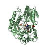
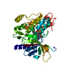
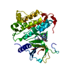
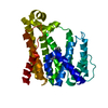
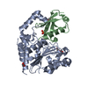
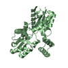
| Title | Crystal structure of the ES2 intermediate form of human hydroxymethylbilane synthase | ||||||||||||
 Components Components | Porphobilinogen deaminase | ||||||||||||
 Keywords Keywords | TRANSFERASE / Porphyrin synthesis | ||||||||||||
| Function / homology |  Function and homology information Function and homology informationhydroxymethylbilane synthase / hydroxymethylbilane synthase activity / heme A biosynthetic process / heme B biosynthetic process / : / Heme biosynthesis / heme biosynthetic process / cytoplasm / cytosol Similarity search - Function | ||||||||||||
| Biological species |  Homo sapiens (human) Homo sapiens (human) | ||||||||||||
| Method |  X-RAY DIFFRACTION / SYNCHROTRON / MOLECULAR REPLACEMENT / Resolution: 1.79 Å X-RAY DIFFRACTION / SYNCHROTRON / MOLECULAR REPLACEMENT / Resolution: 1.79 Å | ||||||||||||
 Authors Authors | Sato, H. / Sugishima, M. / Wada, K. / Hirabayashi, K. / Tsukaguchi, M. | ||||||||||||
| Funding support |  Japan, 3items Japan, 3items
| ||||||||||||
 Citation Citation | Journal: Biochem.J. / Year: 2021 Title: Crystal structures of hydroxymethylbilane synthase complexed with a substrate analog: a single substrate-binding site for four consecutive condensation steps. Authors: Sato, H. / Sugishima, M. / Tsukaguchi, M. / Masuko, T. / Iijima, M. / Takano, M. / Omata, Y. / Hirabayashi, K. / Wada, K. / Hisaeda, Y. / Yamamoto, K. | ||||||||||||
| History |
|
- Structure visualization
| Structure viewer | Molecule: MolmilJmol/JSmol |
|---|
- Downloads & links
-Download
| PDBx/mmCIF format | 7ccz.cif.gz | 161.2 KB | Display | PDBx/mmCIF format |
|---|---|---|---|---|
| PDB format | pdb7ccz.ent.gz | 114.4 KB | Display | PDB format |
| PDBx/mmJSON format | 7ccz.json.gz | Tree view | PDBx/mmJSON format | |
| Others |  Other downloads Other downloads |
-Validation report
| Arichive directory | https://data.pdbj.org/pub/pdb/validation_reports/cc/7cczftp://data.pdbj.org/pub/pdb/validation_reports/cc/7ccz | HTTPS FTP |
|---|
-Related structure data
| Related structure data |  7ccxC  7ccyC  7cd0C  3ecrS S: Starting model for refinement C: citing same article ( |
|---|---|
| Similar structure data |


-Links
 PDBj
PDBj

- Assembly
| Deposited unit | 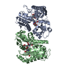
| ||||||||||||
|---|---|---|---|---|---|---|---|---|---|---|---|---|---|
| 1 | 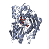
| ||||||||||||
| 2 | 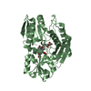
| ||||||||||||
| Unit cell |
|
-Components
| #1: Protein | Mass: 39383.012 Da / Num. of mol.: 2 Source method: isolated from a genetically manipulated source Source: (gene. exp.) Homo sapiens (human) / Gene: HMBS, PBGD, UPS / Production host:  #2: Chemical |   Mass: 838.810 Da / Num. of mol.: 2 / Source method: obtained synthetically / Formula: C40H46N4O16 / Feature type: SUBJECT OF INVESTIGATION Mass: 838.810 Da / Num. of mol.: 2 / Source method: obtained synthetically / Formula: C40H46N4O16 / Feature type: SUBJECT OF INVESTIGATION#3: Water | ChemComp-HOH / |  Mass: 18.015 Da / Num. of mol.: 366 / Source method: isolated from a natural source / Formula: H2O Mass: 18.015 Da / Num. of mol.: 366 / Source method: isolated from a natural source / Formula: H2OHas ligand of interest | Y | Has protein modification | Y | |
|---|
-Experimental details
-Experiment
| Experiment | Method: X-RAY DIFFRACTION / Number of used crystals: 1 |
|---|
- Sample preparation
| Crystal | Density Matthews: 2.23 Å3/Da / Density % sol: 44.96 % |
|---|---|
| Crystal grow | Temperature: 293 K / Method: vapor diffusion, sitting drop / pH: 8.3 Details: PEG 3350, diammonium hydrogen citrate, dithiothreitol |
-Data collection
| Diffraction | Mean temperature: 100 K / Serial crystal experiment: N |
|---|---|
| Diffraction source | Source: SYNCHROTRON / Site: SPring-8 / Beamline: BL44XU / Wavelength: 0.9 Å |
| Detector | Type: RAYONIX MX225HE / Detector: CCD / Date: Nov 10, 2017 |
| Radiation | Protocol: SINGLE WAVELENGTH / Monochromatic (M) / Laue (L): M / Scattering type: x-ray |
| Radiation wavelength | Wavelength: 0.9 Å / Relative weight: 1 |
| Reflection | Resolution: 1.79→45.2 Å / Num. obs: 66546 / % possible obs: 99.8 % / Redundancy: 3.4 % / Biso Wilson estimate: 27.88 Å2 / CC1/2: 0.998 / Rsym value: 0.072 / Net I/σ(I): 9.33 |
| Reflection shell | Resolution: 1.79→1.9 Å / Redundancy: 3.4 % / Mean I/σ(I) obs: 1.3 / Num. unique obs: 20529 / CC1/2: 0.688 / Rsym value: 1.081 / % possible all: 99.2 |
- Processing
| Software |
| ||||||||||||||||||||||||||||||||||||||||||||||||||||||||||||||||||||||||||||||||||||||||||||||||||||||||||||||||||||||||||||||||||||||||||||||||||||||||||||||||||||||||
|---|---|---|---|---|---|---|---|---|---|---|---|---|---|---|---|---|---|---|---|---|---|---|---|---|---|---|---|---|---|---|---|---|---|---|---|---|---|---|---|---|---|---|---|---|---|---|---|---|---|---|---|---|---|---|---|---|---|---|---|---|---|---|---|---|---|---|---|---|---|---|---|---|---|---|---|---|---|---|---|---|---|---|---|---|---|---|---|---|---|---|---|---|---|---|---|---|---|---|---|---|---|---|---|---|---|---|---|---|---|---|---|---|---|---|---|---|---|---|---|---|---|---|---|---|---|---|---|---|---|---|---|---|---|---|---|---|---|---|---|---|---|---|---|---|---|---|---|---|---|---|---|---|---|---|---|---|---|---|---|---|---|---|---|---|---|---|---|---|---|
| Refinement | Method to determine structure: MOLECULAR REPLACEMENT Starting model: 3ECR Resolution: 1.79→44.87 Å / SU ML: 0.2453 / Cross valid method: FREE R-VALUE / σ(F): 1.36 / Phase error: 27.4229 Stereochemistry target values: GeoStd + Monomer Library + CDL v1.2
| ||||||||||||||||||||||||||||||||||||||||||||||||||||||||||||||||||||||||||||||||||||||||||||||||||||||||||||||||||||||||||||||||||||||||||||||||||||||||||||||||||||||||
| Solvent computation | Shrinkage radii: 0.9 Å / VDW probe radii: 1.11 Å / Solvent model: FLAT BULK SOLVENT MODEL | ||||||||||||||||||||||||||||||||||||||||||||||||||||||||||||||||||||||||||||||||||||||||||||||||||||||||||||||||||||||||||||||||||||||||||||||||||||||||||||||||||||||||
| Displacement parameters | Biso mean: 34.85 Å2 | ||||||||||||||||||||||||||||||||||||||||||||||||||||||||||||||||||||||||||||||||||||||||||||||||||||||||||||||||||||||||||||||||||||||||||||||||||||||||||||||||||||||||
| Refinement step | Cycle: LAST / Resolution: 1.79→44.87 Å
| ||||||||||||||||||||||||||||||||||||||||||||||||||||||||||||||||||||||||||||||||||||||||||||||||||||||||||||||||||||||||||||||||||||||||||||||||||||||||||||||||||||||||
| Refine LS restraints |
| ||||||||||||||||||||||||||||||||||||||||||||||||||||||||||||||||||||||||||||||||||||||||||||||||||||||||||||||||||||||||||||||||||||||||||||||||||||||||||||||||||||||||
| LS refinement shell |
|
