 Movie
Movie Controller

[English] 日本語
 Yorodumi
- PDB-3bzs: Crystal structure of EscU C-terminal domain with N262D mutation, ... -
+ Open data

- Basic information
| Entry | Database: PDB / ID: 3bzs | ||||||
|---|---|---|---|---|---|---|---|
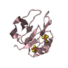
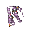
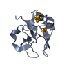
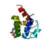
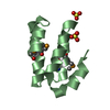
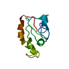
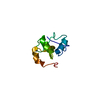
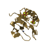
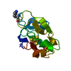
| Title | Crystal structure of EscU C-terminal domain with N262D mutation, Space group P 21 21 21 | ||||||
 Components Components | EscU | ||||||
 Keywords Keywords | MEMBRANE PROTEIN / PROTEIN TRANSPORT / Auto cleavage protein / intein / T3SS / TTSS / asparagine cyclization / membrane | ||||||
| Function / homology |  Function and homology information Function and homology informationdioxygenase activity / protein secretion / isomerase activity / plasma membrane Similarity search - Function | ||||||
| Biological species |  | ||||||
| Method |  X-RAY DIFFRACTION / Resolution: 1.48 Å X-RAY DIFFRACTION / Resolution: 1.48 Å | ||||||
 Authors Authors | Zarivach, R. / Deng, W. / Vuckovic, M. / Felise, H.B. / Nguyen, H.V. / Miller, S.I. / Finlay, B.B. / Strynadka, N.C.J. | ||||||
 Citation Citation | Journal: Nature / Year: 2008 Title: Structural analysis of the essential self-cleaving type III secretion proteins EscU and SpaS. Authors: Zarivach, R. / Deng, W. / Vuckovic, M. / Felise, H.B. / Nguyen, H.V. / Miller, S.I. / Finlay, B.B. / Strynadka, N.C. | ||||||
| History |
|
- Structure visualization
| Structure viewer | Molecule: MolmilJmol/JSmol |
|---|
- Downloads & links
-Download
| PDBx/mmCIF format | 3bzs.cif.gz | 34.8 KB | Display | PDBx/mmCIF format |
|---|---|---|---|---|
| PDB format | pdb3bzs.ent.gz | 23.4 KB | Display | PDB format |
| PDBx/mmJSON format | 3bzs.json.gz | Tree view | PDBx/mmJSON format | |
| Others |  Other downloads Other downloads |
-Validation report
| Arichive directory | https://data.pdbj.org/pub/pdb/validation_reports/bz/3bzsftp://data.pdbj.org/pub/pdb/validation_reports/bz/3bzs | HTTPS FTP |
|---|
-Related structure data
| Related structure data | 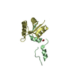 3bzlC 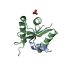 3bzoC 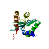 3bzpC  3bzrC  3bztC  3bzvC 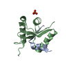 3bzxC 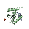 3bzyC 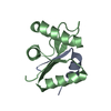 3bzzC 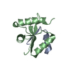 3c00C 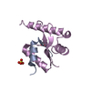 3c01C  3c03C C: citing same article ( |
|---|---|
| Similar structure data |

-Links
 PDBj
PDBj
- Assembly
| Deposited unit | 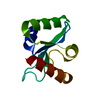
| ||||||||
|---|---|---|---|---|---|---|---|---|---|
| 1 |
| ||||||||
| Unit cell |
|
-Components
| #1: Protein | Mass: 15454.796 Da / Num. of mol.: 1 / Fragment: C-terminal domain / Mutation: N262D Source method: isolated from a genetically manipulated source Source: (gene. exp.) |
|---|---|
| #2: Water | ChemComp-HOH /  Mass: 18.015 Da / Num. of mol.: 96 / Source method: isolated from a natural source / Formula: H2O Mass: 18.015 Da / Num. of mol.: 96 / Source method: isolated from a natural source / Formula: H2O |
-Experimental details
-Experiment
| Experiment | Method: X-RAY DIFFRACTION / Number of used crystals: 1 |
|---|
- Sample preparation
| Crystal | Density Matthews: 1.48 Å3/Da / Density % sol: 17.31 % |
|---|---|
| Crystal grow | Temperature: 293 K / Method: microbatch / pH: 6.5 Details: PEG 3350, NaCl, Bis-Tris, pH 6.5, Microbatch, temperature 293K |
-Data collection
| Diffraction | Mean temperature: 100 K | |||||||||||||||||||||||||||||||||||||||||||||||||||||||||||||||||||||||||||||
|---|---|---|---|---|---|---|---|---|---|---|---|---|---|---|---|---|---|---|---|---|---|---|---|---|---|---|---|---|---|---|---|---|---|---|---|---|---|---|---|---|---|---|---|---|---|---|---|---|---|---|---|---|---|---|---|---|---|---|---|---|---|---|---|---|---|---|---|---|---|---|---|---|---|---|---|---|---|---|
| Diffraction source | Source: ROTATING ANODE / Type: RIGAKU MICROMAX-007 HF / Wavelength: 1.5418 Å | |||||||||||||||||||||||||||||||||||||||||||||||||||||||||||||||||||||||||||||
| Detector | Type: MAR scanner 345 mm plate / Detector: IMAGE PLATE | |||||||||||||||||||||||||||||||||||||||||||||||||||||||||||||||||||||||||||||
| Radiation | Protocol: SINGLE WAVELENGTH / Monochromatic (M) / Laue (L): M / Scattering type: x-ray | |||||||||||||||||||||||||||||||||||||||||||||||||||||||||||||||||||||||||||||
| Radiation wavelength | Wavelength: 1.5418 Å / Relative weight: 1 | |||||||||||||||||||||||||||||||||||||||||||||||||||||||||||||||||||||||||||||
| Reflection | Resolution: 1.48→50 Å / Num. all: 16014 / Num. obs: 14461 / % possible obs: 90.3 % / Observed criterion σ(F): 2 / Observed criterion σ(I): 2 / Redundancy: 9.7 % / Rmerge(I) obs: 0.053 / Χ2: 1.573 / Net I/σ(I): 14.3 | |||||||||||||||||||||||||||||||||||||||||||||||||||||||||||||||||||||||||||||
| Reflection shell |
|
- Processing
| Software |
| |||||||||||||||||||||||||||||||||||||||||||||||||||||||||||||||||||||||||||||||||||||||||||||||||||||||||||||||||||||||||||||||||||||||||||||||||||||||||||||||||||||||||||||||
|---|---|---|---|---|---|---|---|---|---|---|---|---|---|---|---|---|---|---|---|---|---|---|---|---|---|---|---|---|---|---|---|---|---|---|---|---|---|---|---|---|---|---|---|---|---|---|---|---|---|---|---|---|---|---|---|---|---|---|---|---|---|---|---|---|---|---|---|---|---|---|---|---|---|---|---|---|---|---|---|---|---|---|---|---|---|---|---|---|---|---|---|---|---|---|---|---|---|---|---|---|---|---|---|---|---|---|---|---|---|---|---|---|---|---|---|---|---|---|---|---|---|---|---|---|---|---|---|---|---|---|---|---|---|---|---|---|---|---|---|---|---|---|---|---|---|---|---|---|---|---|---|---|---|---|---|---|---|---|---|---|---|---|---|---|---|---|---|---|---|---|---|---|---|---|---|---|
| Refinement | Resolution: 1.48→37.8 Å / Cor.coef. Fo:Fc: 0.957 / Cor.coef. Fo:Fc free: 0.949 / SU B: 2.666 / SU ML: 0.051 / TLS residual ADP flag: LIKELY RESIDUAL / Cross valid method: THROUGHOUT / σ(F): 0 / ESU R: 0.097 / ESU R Free: 0.091 / Stereochemistry target values: MAXIMUM LIKELIHOOD / Details: HYDROGENS HAVE BEEN ADDED IN THE RIDING POSITIONS
| |||||||||||||||||||||||||||||||||||||||||||||||||||||||||||||||||||||||||||||||||||||||||||||||||||||||||||||||||||||||||||||||||||||||||||||||||||||||||||||||||||||||||||||||
| Solvent computation | Ion probe radii: 0.8 Å / Shrinkage radii: 0.8 Å / VDW probe radii: 1.4 Å / Solvent model: MASK | |||||||||||||||||||||||||||||||||||||||||||||||||||||||||||||||||||||||||||||||||||||||||||||||||||||||||||||||||||||||||||||||||||||||||||||||||||||||||||||||||||||||||||||||
| Displacement parameters | Biso mean: 12.043 Å2
| |||||||||||||||||||||||||||||||||||||||||||||||||||||||||||||||||||||||||||||||||||||||||||||||||||||||||||||||||||||||||||||||||||||||||||||||||||||||||||||||||||||||||||||||
| Refinement step | Cycle: LAST / Resolution: 1.48→37.8 Å
| |||||||||||||||||||||||||||||||||||||||||||||||||||||||||||||||||||||||||||||||||||||||||||||||||||||||||||||||||||||||||||||||||||||||||||||||||||||||||||||||||||||||||||||||
| Refine LS restraints |
| |||||||||||||||||||||||||||||||||||||||||||||||||||||||||||||||||||||||||||||||||||||||||||||||||||||||||||||||||||||||||||||||||||||||||||||||||||||||||||||||||||||||||||||||
| LS refinement shell | Resolution: 1.48→1.521 Å / Total num. of bins used: 20
| |||||||||||||||||||||||||||||||||||||||||||||||||||||||||||||||||||||||||||||||||||||||||||||||||||||||||||||||||||||||||||||||||||||||||||||||||||||||||||||||||||||||||||||||
| Refinement TLS params. | Method: refined / Refine-ID: X-RAY DIFFRACTION
| |||||||||||||||||||||||||||||||||||||||||||||||||||||||||||||||||||||||||||||||||||||||||||||||||||||||||||||||||||||||||||||||||||||||||||||||||||||||||||||||||||||||||||||||
| Refinement TLS group |
|
