 Movie
Movie Controller

[English] 日本語
 Yorodumi
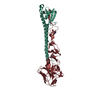
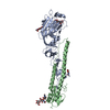
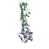
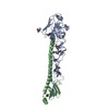
- PDB-3al4: Crystal structure of the swine-origin A (H1N1)-2009 influenza A v... -
+ Open data

- Basic information
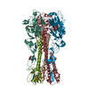
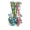
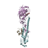
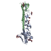
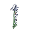
| Entry | Database: PDB / ID: 3al4 | ||||||||||||
|---|---|---|---|---|---|---|---|---|---|---|---|---|---|
| Title | Crystal structure of the swine-origin A (H1N1)-2009 influenza A virus hemagglutinin (HA) reveals similar antigenicity to that of the 1918 pandemic virus | ||||||||||||
 Components Components | (Hemagglutinin) x 2 | ||||||||||||
 Keywords Keywords | VIRAL PROTEIN/VIRAL PROTEIN / TRIMER / ENVELOPE PROTEIN / HEMAGGLUTININ / VIRAL PROTEIN-VIRAL PROTEIN complex | ||||||||||||
| Function / homology |  Function and homology information Function and homology informationviral budding from plasma membrane / clathrin-dependent endocytosis of virus by host cell / host cell surface receptor binding / fusion of virus membrane with host plasma membrane / fusion of virus membrane with host endosome membrane / viral envelope / virion attachment to host cell / host cell plasma membrane / virion membrane / membrane / metal ion binding Similarity search - Function | ||||||||||||
| Biological species |   Influenza A virus Influenza A virus | ||||||||||||
| Method |  X-RAY DIFFRACTION / SYNCHROTRON / MOLECULAR REPLACEMENT / Resolution: 2.872 Å X-RAY DIFFRACTION / SYNCHROTRON / MOLECULAR REPLACEMENT / Resolution: 2.872 Å | ||||||||||||
 Authors Authors | Zhang, W. / Qi, J.X. / Shi, Y. / Li, Q. / Yan, J.H. / Gao, G.F. | ||||||||||||
 Citation Citation | Journal: Protein Cell / Year: 2010 Title: Crystal structure of the swine-origin A (H1N1)-2009 influenza A virus hemagglutinin (HA) reveals similar antigenicity to that of the 1918 pandemic virus Authors: Zhang, W. / Qi, J. / Shi, Y. / Li, Q. / Gao, F. / Sun, Y. / Lu, X. / Lu, Q. / Vavricka, C.J. / Liu, D. / Yan, J. / Gao, G.F. | ||||||||||||
| History |
|
- Structure visualization
| Structure viewer | Molecule: MolmilJmol/JSmol |
|---|
- Downloads & links
-Download
| PDBx/mmCIF format | 3al4.cif.gz | 1.1 MB | Display | PDBx/mmCIF format |
|---|---|---|---|---|
| PDB format | pdb3al4.ent.gz | 984.7 KB | Display | PDB format |
| PDBx/mmJSON format | 3al4.json.gz | Tree view | PDBx/mmJSON format | |
| Others |  Other downloads Other downloads |
-Validation report
| Arichive directory | https://data.pdbj.org/pub/pdb/validation_reports/al/3al4ftp://data.pdbj.org/pub/pdb/validation_reports/al/3al4 | HTTPS FTP |
|---|
-Related structure data
| Related structure data | 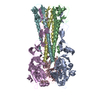 1ru7S S: Starting model for refinement |
|---|---|
| Similar structure data |
-Links
 PDBj
PDBj


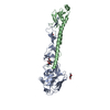
- Assembly
| Deposited unit | 
| ||||||||
|---|---|---|---|---|---|---|---|---|---|
| 1 | 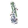
| ||||||||
| 2 | 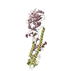
| ||||||||
| 3 | 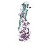
| ||||||||
| 4 | 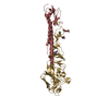
| ||||||||
| 5 | 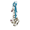
| ||||||||
| 6 | 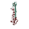
| ||||||||
| Unit cell |
|
-Components
-Protein , 2 types, 12 molecules ACEGIKBDFHJL
| #1: Protein | Mass: 36989.715 Da / Num. of mol.: 6 / Fragment: UNP residues 18-344 Source method: isolated from a genetically manipulated source Details: HEMAGGLUTININ HA1 / Source: (gene. exp.) Influenza A virus / Strain: A/CALIFORNIA/04/2009(H1N1) / Gene: HEMAGGLUTININ / Production host:   Spodoptera frugiperda (fall armyworm) / Strain (production host): SF9 / References: UniProt: C3W5S1 Spodoptera frugiperda (fall armyworm) / Strain (production host): SF9 / References: UniProt: C3W5S1#2: Protein | Mass: 20757.035 Da / Num. of mol.: 6 / Fragment: UNP residues 345-520 Source method: isolated from a genetically manipulated source Details: HEMAGGLUTININ HA2 / Source: (gene. exp.) Influenza A virus / Strain: A/CALIFORNIA/04/2009(H1N1) / Gene: HEMAGGLUTININ / Production host: Spodoptera frugiperda (fall armyworm) / Strain (production host): SF9 / References: UniProt: C3W5S1 |
|---|
-Sugars , 3 types, 23 molecules 
| #3: Polysaccharide | 2-acetamido-2-deoxy-beta-D-glucopyranose-(1-4)-2-acetamido-2-deoxy-beta-D-glucopyranose Source method: isolated from a genetically manipulated source #4: Polysaccharide | Source method: isolated from a genetically manipulated source #5: Sugar | ChemComp-NAG /  Type: D-saccharide, beta linking / Mass: 221.208 Da / Num. of mol.: 16 Type: D-saccharide, beta linking / Mass: 221.208 Da / Num. of mol.: 16Source method: isolated from a genetically manipulated source Formula: C8H15NO6 |
|---|
-Non-polymers , 1 types, 317 molecules 
| #6: Water | ChemComp-HOH / Mass: 18.015 Da / Num. of mol.: 317 / Source method: isolated from a natural source / Formula: H2O |
|---|
-Details
| Has protein modification | Y |
|---|
-Experimental details
-Experiment
| Experiment | Method: X-RAY DIFFRACTION / Number of used crystals: 1 |
|---|
- Sample preparation
| Crystal | Density Matthews: 2.18 Å3/Da / Density % sol: 43.59 % |
|---|---|
| Crystal grow | Temperature: 291 K / Method: vapor diffusion, hanging drop / pH: 6.5 Details: 10% PEG 6000, 5% MPD, 0.1M MES, PH 6.5, VAPOR DIFFUSION, HANGING DROP, temperature 291K |
-Data collection
| Diffraction | Mean temperature: 100 K |
|---|---|
| Diffraction source | Source: SYNCHROTRON / Site: SSRF  / Beamline: BL17U / Wavelength: 0.9795 Å / Beamline: BL17U / Wavelength: 0.9795 Å |
| Detector | Type: MARMOSAIC 225 mm CCD / Detector: CCD / Date: Jan 23, 2010 / Details: MIRRORS |
| Radiation | Monochromator: GRAPHITE / Protocol: SINGLE WAVELENGTH / Monochromatic (M) / Laue (L): M / Scattering type: x-ray |
| Radiation wavelength | Wavelength: 0.9795 Å / Relative weight: 1 |
| Reflection | Resolution: 2.872→50 Å / Num. obs: 64796 / % possible obs: 98 % / Observed criterion σ(I): 1.5 / Redundancy: 3.7 % / Biso Wilson estimate: 56.27 Å2 / Rmerge(I) obs: 0.105 / Rsym value: 0.105 / Net I/σ(I): 12.9 |
| Reflection shell | Resolution: 2.87→3 Å / Redundancy: 2.7 % / Rmerge(I) obs: 0.501 / Mean I/σ(I) obs: 1.9 / Rsym value: 0.501 / % possible all: 90.7 |
- Processing
| Software |
| |||||||||||||||||||||||||||||||||||||||||||||||||||||||||||||||||||||||||||||||||||||||||||||||||||||||||||||||||||||||||||||||||||||||||||||||||||||||||||||||||
|---|---|---|---|---|---|---|---|---|---|---|---|---|---|---|---|---|---|---|---|---|---|---|---|---|---|---|---|---|---|---|---|---|---|---|---|---|---|---|---|---|---|---|---|---|---|---|---|---|---|---|---|---|---|---|---|---|---|---|---|---|---|---|---|---|---|---|---|---|---|---|---|---|---|---|---|---|---|---|---|---|---|---|---|---|---|---|---|---|---|---|---|---|---|---|---|---|---|---|---|---|---|---|---|---|---|---|---|---|---|---|---|---|---|---|---|---|---|---|---|---|---|---|---|---|---|---|---|---|---|---|---|---|---|---|---|---|---|---|---|---|---|---|---|---|---|---|---|---|---|---|---|---|---|---|---|---|---|---|---|---|---|---|
| Refinement | Method to determine structure: MOLECULAR REPLACEMENT Starting model: PDB ENTRY 1RU7 Resolution: 2.872→24.19 Å / Occupancy max: 1 / Occupancy min: 1 / SU ML: 0.37 / σ(F): 0.07 / Phase error: 29.51 / Stereochemistry target values: ML
| |||||||||||||||||||||||||||||||||||||||||||||||||||||||||||||||||||||||||||||||||||||||||||||||||||||||||||||||||||||||||||||||||||||||||||||||||||||||||||||||||
| Solvent computation | Shrinkage radii: 0.9 Å / VDW probe radii: 1.11 Å / Solvent model: FLAT BULK SOLVENT MODEL / Bsol: 39.94 Å2 / ksol: 0.29 e/Å3 | |||||||||||||||||||||||||||||||||||||||||||||||||||||||||||||||||||||||||||||||||||||||||||||||||||||||||||||||||||||||||||||||||||||||||||||||||||||||||||||||||
| Displacement parameters | Biso max: 330.34 Å2 / Biso mean: 81.17 Å2 / Biso min: 16.64 Å2
| |||||||||||||||||||||||||||||||||||||||||||||||||||||||||||||||||||||||||||||||||||||||||||||||||||||||||||||||||||||||||||||||||||||||||||||||||||||||||||||||||
| Refinement step | Cycle: LAST / Resolution: 2.872→24.19 Å
| |||||||||||||||||||||||||||||||||||||||||||||||||||||||||||||||||||||||||||||||||||||||||||||||||||||||||||||||||||||||||||||||||||||||||||||||||||||||||||||||||
| Refine LS restraints |
| |||||||||||||||||||||||||||||||||||||||||||||||||||||||||||||||||||||||||||||||||||||||||||||||||||||||||||||||||||||||||||||||||||||||||||||||||||||||||||||||||
| LS refinement shell |
| |||||||||||||||||||||||||||||||||||||||||||||||||||||||||||||||||||||||||||||||||||||||||||||||||||||||||||||||||||||||||||||||||||||||||||||||||||||||||||||||||
| Refinement TLS params. | Method: refined / Origin x: -20.2272 Å / Origin y: -28.1217 Å / Origin z: -32.6646 Å
| |||||||||||||||||||||||||||||||||||||||||||||||||||||||||||||||||||||||||||||||||||||||||||||||||||||||||||||||||||||||||||||||||||||||||||||||||||||||||||||||||
| Refinement TLS group |
|
