 Movie
Movie Controller


+ Open data

- Basic information
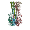
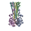
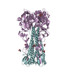
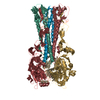
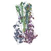
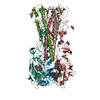
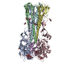
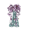
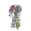
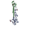
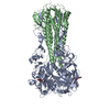
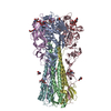
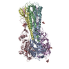
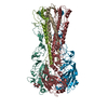
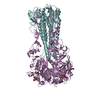
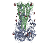
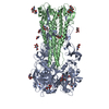
| Entry | Database: PDB / ID: 1ruz | |||||||||
|---|---|---|---|---|---|---|---|---|---|---|
| Title | 1918 H1 Hemagglutinin | |||||||||
 Components Components | (hemagglutinin) x 2 | |||||||||
 Keywords Keywords | VIRAL PROTEIN / Hemagglutinin / INFLUENZA A VIRUS | |||||||||
| Function / homology |  Function and homology information Function and homology informationviral budding from plasma membrane / clathrin-dependent endocytosis of virus by host cell / host cell surface receptor binding / fusion of virus membrane with host plasma membrane / fusion of virus membrane with host endosome membrane / viral envelope / virion attachment to host cell / host cell plasma membrane / virion membrane / membrane Similarity search - Function | |||||||||
| Biological species |   Influenza A virus Influenza A virus | |||||||||
| Method |  X-RAY DIFFRACTION / SYNCHROTRON / MOLECULAR REPLACEMENT / Resolution: 2.9 Å X-RAY DIFFRACTION / SYNCHROTRON / MOLECULAR REPLACEMENT / Resolution: 2.9 Å | |||||||||
 Authors Authors | Skehel, J.J. / Gamblin, S.J. / Haire, L.F. / Russell, R.J. / Stevens, D.J. / Xiao, B. / Ha, Y. / Vasisht, N. / Steinhauer, D.A. / Daniels, R.S. | |||||||||
 Citation Citation | Journal: Science / Year: 2004 Title: The structure and receptor binding properties of the 1918 influenza hemagglutinin. Authors: Gamblin, S.J. / Haire, L.F. / Russell, R.J. / Stevens, D.J. / Xiao, B. / Ha, Y. / Vasisht, N. / Steinhauer, D.A. / Daniels, R.S. / Elliot, A. / Wiley, D.C. / Skehel, J.J. | |||||||||
| History |
|
- Structure visualization
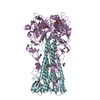
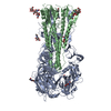
| Structure viewer | Molecule: MolmilJmol/JSmol |
|---|
- Downloads & links
-Download
| PDBx/mmCIF format | 1ruz.cif.gz | 304.7 KB | Display | PDBx/mmCIF format |
|---|---|---|---|---|
| PDB format | pdb1ruz.ent.gz | 248.1 KB | Display | PDB format |
| PDBx/mmJSON format | 1ruz.json.gz | Tree view | PDBx/mmJSON format | |
| Others |  Other downloads Other downloads |
-Validation report
| Arichive directory | https://data.pdbj.org/pub/pdb/validation_reports/ru/1ruzftp://data.pdbj.org/pub/pdb/validation_reports/ru/1ruz | HTTPS FTP |
|---|
-Related structure data
| Related structure data |  1ru7SC 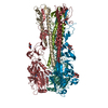 1ruyC  1rv0C 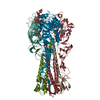 1rvtC 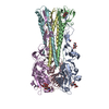 1rvxC  1rvzC S: Starting model for refinement C: citing same article ( |
|---|---|
| Similar structure data |

-Links
 PDBj
PDBj


- Assembly
| Deposited unit | 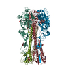
| ||||||||
|---|---|---|---|---|---|---|---|---|---|
| 1 |
| ||||||||
| Unit cell |
|
-Components
| #1: Protein | Mass: 36125.465 Da / Num. of mol.: 3 / Source method: isolated from a natural source / Details: Hemagglutinin HA1 chain Source: (natural) Influenza A virus (A/South Carolina/1/18 (H1N1))Genus: Influenzavirus A / Cell: Vaccinia virus infected CV1 cells / Species: Influenza A virus / Strain: A-SOUTH CAROLINA-1-18 (H1N1) / References: UniProt: Q9WFZ1, UniProt: Q9WFX3*PLUS #2: Protein | Mass: 18212.092 Da / Num. of mol.: 3 / Source method: isolated from a natural source / Details: Hemagglutinin HA2 chain Source: (natural) Influenza A virus (A/South Carolina/1/18 (H1N1))Genus: Influenzavirus A / Cell: Vaccinia virus infected CV1 cells / Species: Influenza A virus / Strain: A-SOUTH CAROLINA-1-18 (H1N1) / References: UniProt: Q9WFZ1, UniProt: Q9WFX3*PLUS #3: Sugar | ChemComp-NDG /   Type: D-saccharide, alpha linking / Mass: 221.208 Da / Num. of mol.: 9 Type: D-saccharide, alpha linking / Mass: 221.208 Da / Num. of mol.: 9Source method: isolated from a genetically manipulated source Formula: C8H15NO6 #4: Sugar |   Type: D-saccharide, beta linking / Mass: 221.208 Da / Num. of mol.: 2 Type: D-saccharide, beta linking / Mass: 221.208 Da / Num. of mol.: 2Source method: isolated from a genetically manipulated source Formula: C8H15NO6 #5: Water | ChemComp-HOH / |  Mass: 18.015 Da / Num. of mol.: 549 / Source method: isolated from a natural source / Formula: H2O Mass: 18.015 Da / Num. of mol.: 549 / Source method: isolated from a natural source / Formula: H2OHas protein modification | Y | |
|---|
-Experimental details
-Experiment
| Experiment | Method: X-RAY DIFFRACTION / Number of used crystals: 1 |
|---|
- Sample preparation
| Crystal | Density Matthews: 3.46 Å3/Da / Density % sol: 64.43 % |
|---|---|
| Crystal grow | Temperature: 291 K / Method: vapor diffusion, hanging drop / pH: 6.5 Details: PEG3350, TrisHCl, Bis tris propane, pH 6.5, VAPOR DIFFUSION, HANGING DROP, temperature 291K |
-Data collection
| Diffraction | Mean temperature: 100 K |
|---|---|
| Diffraction source | Source: SYNCHROTRON / Site: SRS  / Beamline: PX14.2 / Beamline: PX14.2 |
| Detector | Type: ADSC QUANTUM 4 / Detector: CCD / Date: Jun 20, 2003 |
| Radiation | Protocol: SINGLE WAVELENGTH / Monochromatic (M) / Laue (L): M / Scattering type: x-ray |
| Radiation wavelength | Relative weight: 1 |
| Reflection | Resolution: 2.9→20 Å / Num. all: 51196 / Num. obs: 49134 / % possible obs: 96.1 % / Observed criterion σ(F): 0 / Observed criterion σ(I): 0 / Redundancy: 19 % / Rmerge(I) obs: 0.085 / Net I/σ(I): 11.9 |
| Reflection shell | Resolution: 2.9→3.1 Å / Rmerge(I) obs: 0.574 / Mean I/σ(I) obs: 1.8 / % possible all: 88.4 |
- Processing
| Software |
| ||||||||||||||||||||
|---|---|---|---|---|---|---|---|---|---|---|---|---|---|---|---|---|---|---|---|---|---|
| Refinement | Method to determine structure: MOLECULAR REPLACEMENT Starting model: PDB entry 1RU7 Resolution: 2.9→20 Å / σ(F): 0 / Stereochemistry target values: Engh & Huber
| ||||||||||||||||||||
| Refinement step | Cycle: LAST / Resolution: 2.9→20 Å
|
