 Movie
Movie Controller


+ Open data

- Basic information
| Entry | Database: PDB / ID: 2wrg | |||||||||
|---|---|---|---|---|---|---|---|---|---|---|
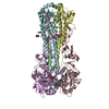
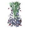
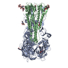
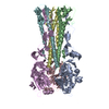
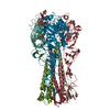
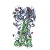
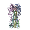
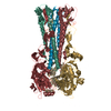
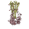
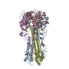
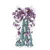
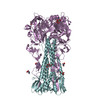
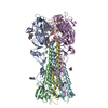
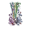
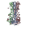
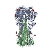
| Title | structure of H1 1918 hemagglutinin with human receptor | |||||||||
 Components Components |
| |||||||||
 Keywords Keywords | VIRAL PROTEIN / ENVELOPE PROTEIN / GLYCOPROTEIN / LIPOPROTEIN | |||||||||
| Function / homology |  Function and homology information Function and homology informationviral budding from plasma membrane / clathrin-dependent endocytosis of virus by host cell / host cell surface receptor binding / fusion of virus membrane with host plasma membrane / fusion of virus membrane with host endosome membrane / viral envelope / virion attachment to host cell / host cell plasma membrane / virion membrane / membrane Similarity search - Function | |||||||||
| Biological species |   INFLUENZA A VIRUS INFLUENZA A VIRUS | |||||||||
| Method |  X-RAY DIFFRACTION / OTHER / Resolution: 3 Å X-RAY DIFFRACTION / OTHER / Resolution: 3 Å | |||||||||
 Authors Authors | Liu, J. / Stevens, D.J. / Haire, L.F. / Walker, P.A. / Coombs, P.J. / Russell, R.J. / Gamblin, S.J. / Skehel, J.J. | |||||||||
 Citation Citation | Journal: Proc.Natl.Acad.Sci.USA / Year: 2009 Title: From the Cover: Structures of Receptor Complexes Formed by Hemagglutinins from the Asian Influenza Pandemic of 1957. Authors: Liu, J. / Stevens, D.J. / Haire, L.F. / Walker, P.A. / Coombs, P.J. / Russell, R.J. / Gamblin, S.J. / Skehel, J.J. | |||||||||
| History |
|
- Structure visualization
| Structure viewer | Molecule: MolmilJmol/JSmol |
|---|
- Downloads & links
-Download
| PDBx/mmCIF format | 2wrg.cif.gz | 298 KB | Display | PDBx/mmCIF format |
|---|---|---|---|---|
| PDB format | pdb2wrg.ent.gz | 240.6 KB | Display | PDB format |
| PDBx/mmJSON format | 2wrg.json.gz | Tree view | PDBx/mmJSON format | |
| Others |  Other downloads Other downloads |
-Validation report
| Arichive directory | https://data.pdbj.org/pub/pdb/validation_reports/wr/2wrgftp://data.pdbj.org/pub/pdb/validation_reports/wr/2wrg | HTTPS FTP |
|---|
-Related structure data
| Related structure data |  2wr0C 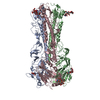 2wr1C 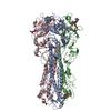 2wr2C  2wr3C  2wr4C  2wr5C  2wr7C  2wrbC  2wrcC 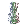 2wrdC  2wreC  2wrfC  2wrhC C: citing same article ( |
|---|---|
| Similar structure data |


-Links
 PDBj
PDBj


- Assembly
| Deposited unit | 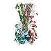
| |||||||||||||||||||||||||||||||||||||||||||||
|---|---|---|---|---|---|---|---|---|---|---|---|---|---|---|---|---|---|---|---|---|---|---|---|---|---|---|---|---|---|---|---|---|---|---|---|---|---|---|---|---|---|---|---|---|---|---|
| 1 |
| |||||||||||||||||||||||||||||||||||||||||||||
| Unit cell |
| |||||||||||||||||||||||||||||||||||||||||||||
| Noncrystallographic symmetry (NCS) | NCS domain:
NCS domain segments:
NCS ensembles :
|
-Components
| #1: Protein | Mass: 35999.309 Da / Num. of mol.: 3 / Fragment: RESIDUES 18-343 / Source method: isolated from a natural source Source: (natural) INFLUENZA A VIRUS (A/BREVIG MISSION/1/1918(H1N1))References: UniProt: Q9WFX3 #2: Protein | Mass: 25047.072 Da / Num. of mol.: 3 / Fragment: RESIDUES 345-566 / Source method: isolated from a natural source Source: (natural) INFLUENZA A VIRUS (A/BREVIG MISSION/1/1918(H1N1))References: UniProt: Q9WFX3 #3: Polysaccharide | N-acetyl-alpha-neuraminic acid-(2-6)-beta-D-galactopyranose-(1-4)-2-acetamido-2-deoxy-beta-D- ...N-acetyl-alpha-neuraminic acid-(2-6)-beta-D-galactopyranose-(1-4)-2-acetamido-2-deoxy-beta-D-glucopyranose-(1-3)-beta-D-galactopyranose | Source method: isolated from a genetically manipulated source #4: Sugar | ChemComp-NAG /   Type: D-saccharide, beta linking / Mass: 221.208 Da / Num. of mol.: 11 Type: D-saccharide, beta linking / Mass: 221.208 Da / Num. of mol.: 11Source method: isolated from a genetically manipulated source Formula: C8H15NO6 Has protein modification | Y | |
|---|
-Experimental details
-Experiment
| Experiment | Method: X-RAY DIFFRACTION |
|---|
- Sample preparation
| Crystal | Density Matthews: 3.54 Å3/Da / Density % sol: 68.9 % / Description: NONE |
|---|
-Data collection
| Diffraction | Mean temperature: 100 K |
|---|---|
| Diffraction source | Source: ROTATING ANODE / Wavelength: 1.5418 |
| Radiation | Protocol: SINGLE WAVELENGTH / Monochromatic (M) / Laue (L): M / Scattering type: x-ray |
| Radiation wavelength | Wavelength: 1.5418 Å / Relative weight: 1 |
| Reflection | Resolution: 3→30 Å / Num. obs: 46270 / % possible obs: 98.5 % / Observed criterion σ(I): 1.3 / Redundancy: 5.1 % / Biso Wilson estimate: 77.87 Å2 / Rmerge(I) obs: 0.16 |
- Processing
| Software | Name: PHENIX / Version: (PHENIX.REFINE) / Classification: refinement | ||||||||||||||||||||||||||||||||||||||||||||||||||||||||||||||||||||||||||||||||||||||||||||||||||||||||||||||||||||||||||||||
|---|---|---|---|---|---|---|---|---|---|---|---|---|---|---|---|---|---|---|---|---|---|---|---|---|---|---|---|---|---|---|---|---|---|---|---|---|---|---|---|---|---|---|---|---|---|---|---|---|---|---|---|---|---|---|---|---|---|---|---|---|---|---|---|---|---|---|---|---|---|---|---|---|---|---|---|---|---|---|---|---|---|---|---|---|---|---|---|---|---|---|---|---|---|---|---|---|---|---|---|---|---|---|---|---|---|---|---|---|---|---|---|---|---|---|---|---|---|---|---|---|---|---|---|---|---|---|---|
| Refinement | Method to determine structure: OTHER Starting model: NONE Resolution: 3→29.68 Å / SU ML: 2.41 / σ(F): 0.02 / Phase error: 30.6 / Stereochemistry target values: MLHL
| ||||||||||||||||||||||||||||||||||||||||||||||||||||||||||||||||||||||||||||||||||||||||||||||||||||||||||||||||||||||||||||||
| Solvent computation | Shrinkage radii: 0.9 Å / VDW probe radii: 1.11 Å / Solvent model: FLAT BULK SOLVENT MODEL / Bsol: 48.06 Å2 / ksol: 0.282 e/Å3 | ||||||||||||||||||||||||||||||||||||||||||||||||||||||||||||||||||||||||||||||||||||||||||||||||||||||||||||||||||||||||||||||
| Displacement parameters |
| ||||||||||||||||||||||||||||||||||||||||||||||||||||||||||||||||||||||||||||||||||||||||||||||||||||||||||||||||||||||||||||||
| Refinement step | Cycle: LAST / Resolution: 3→29.68 Å
| ||||||||||||||||||||||||||||||||||||||||||||||||||||||||||||||||||||||||||||||||||||||||||||||||||||||||||||||||||||||||||||||
| Refine LS restraints |
| ||||||||||||||||||||||||||||||||||||||||||||||||||||||||||||||||||||||||||||||||||||||||||||||||||||||||||||||||||||||||||||||
| Refine LS restraints NCS |
| ||||||||||||||||||||||||||||||||||||||||||||||||||||||||||||||||||||||||||||||||||||||||||||||||||||||||||||||||||||||||||||||
| LS refinement shell |
|
