 Movie
Movie Controller

[English] 日本語
 Yorodumi
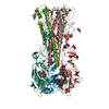
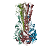
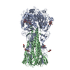
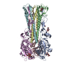
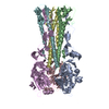
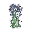
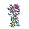
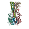
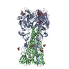
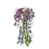
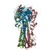
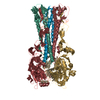
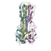
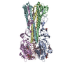
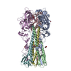
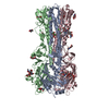
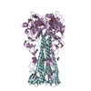
- PDB-3htt: The hemagglutinin structure of an avian H1N1 influenza A virus in... -
+ Open data

- Basic information
| Entry | Database: PDB / ID: 3htt | |||||||||
|---|---|---|---|---|---|---|---|---|---|---|
| Title | The hemagglutinin structure of an avian H1N1 influenza A virus in complex with 2,3-sialyllactose | |||||||||
 Components Components |
| |||||||||
 Keywords Keywords | VIRAL PROTEIN / receptor | |||||||||
| Function / homology |  Function and homology information Function and homology informationclathrin-dependent endocytosis of virus by host cell / host cell surface receptor binding / fusion of virus membrane with host plasma membrane / fusion of virus membrane with host endosome membrane / viral envelope / virion attachment to host cell / host cell plasma membrane Similarity search - Function | |||||||||
| Biological species |   Influenza A virus Influenza A virus | |||||||||
| Method |  X-RAY DIFFRACTION / SYNCHROTRON / MOLECULAR REPLACEMENT / Resolution: 2.95 Å X-RAY DIFFRACTION / SYNCHROTRON / MOLECULAR REPLACEMENT / Resolution: 2.95 Å | |||||||||
 Authors Authors | Wang, G. / Li, A. / Zhang, Q. / Wu, C. / Zhang, R. / Cai, Q. / Song, W. / Yuen, K.-Y. | |||||||||
 Citation Citation | Journal: Virology / Year: 2009 Title: The hemagglutinin structure of an avian H1N1 influenza A virus Authors: Lin, T. / Wang, G. / Li, A. / Zhang, Q. / Wu, C. / Zhang, R. / Cai, Q. / Song, W. / Yuen, K.-Y. | |||||||||
| History |
|
- Structure visualization
| Structure viewer | Molecule: MolmilJmol/JSmol |
|---|
- Downloads & links
-Download
| PDBx/mmCIF format | 3htt.cif.gz | 120.7 KB | Display | PDBx/mmCIF format |
|---|---|---|---|---|
| PDB format | pdb3htt.ent.gz | 91.3 KB | Display | PDB format |
| PDBx/mmJSON format | 3htt.json.gz | Tree view | PDBx/mmJSON format | |
| Others |  Other downloads Other downloads |
-Validation report
| Arichive directory | https://data.pdbj.org/pub/pdb/validation_reports/ht/3httftp://data.pdbj.org/pub/pdb/validation_reports/ht/3htt | HTTPS FTP |
|---|
-Related structure data
| Related structure data |  3htoC  3htpC  3htqC  1ru7S C: citing same article ( S: Starting model for refinement |
|---|---|
| Similar structure data |


-Links
 PDBj
PDBj


- Assembly
| Deposited unit | 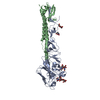
| |||||||||
|---|---|---|---|---|---|---|---|---|---|---|
| 1 | 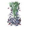
| |||||||||
| Unit cell |
| |||||||||
| Components on special symmetry positions |
|
-Components
-Protein , 2 types, 2 molecules AB
| #1: Protein | Mass: 35946.227 Da / Num. of mol.: 1 / Fragment: HA1 chain, UNP residues 15-338 / Source method: isolated from a natural source Source: (natural) Influenza A virus (A/WDK/JX/12416/2005(H1N1))Strain: WDK/JX/12416/2005 / References: UniProt: C7C6F1 |
|---|---|
| #2: Protein | Mass: 18288.166 Da / Num. of mol.: 1 / Source method: isolated from a natural source Source: (natural) Influenza A virus (A/WDK/JX/12416/2005(H1N1))Strain: WDK/JX/12416/2005 / References: UniProt: C7C6F1*PLUS |
-Sugars , 3 types, 4 molecules 
| #3: Polysaccharide | 2-acetamido-2-deoxy-alpha-D-glucopyranose-(1-4)-2-acetamido-2-deoxy-beta-D-glucopyranose Source method: isolated from a genetically manipulated source |
|---|---|
| #4: Polysaccharide | N-acetyl-alpha-neuraminic acid-(2-3)-beta-D-galactopyranose-(1-4)-alpha-D-glucopyranose / 3'-sialyl-alpha-lactose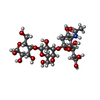  Source method: isolated from a genetically manipulated source Details: oligosaccharide / References: 3'-sialyl-alpha-lactose |
| #5: Sugar |  Type: D-saccharide, beta linking / Mass: 221.208 Da / Num. of mol.: 2 Type: D-saccharide, beta linking / Mass: 221.208 Da / Num. of mol.: 2Source method: isolated from a genetically manipulated source Formula: C8H15NO6 |
-Non-polymers , 1 types, 288 molecules 
| #6: Water | ChemComp-HOH / Mass: 18.015 Da / Num. of mol.: 288 / Source method: isolated from a natural source / Formula: H2O |
|---|
-Details
| Has protein modification | Y | ||
|---|---|---|---|
| Nonpolymer details | THESE THREE SUGERS, SIA3033, GAL3034 AND GLU3035, FORM 2,3-SIALYLLACT| Sequence details | THERE IS NO UNP REFERENCE SEQUENCE DATABASE FOR CHAIN B AT THE TIME OF PROCESSING | |
-Experimental details
-Experiment
| Experiment | Method: X-RAY DIFFRACTION / Number of used crystals: 1 |
|---|
- Sample preparation
| Crystal | Density Matthews: 6.01176 Å3/Da / Density % sol: 79.5401 % Description: the Matthews coefficient is 2.5 and the solvent content is 50.96%, based on their calculations. One monomer per asymmetric unit is used for this calculation. |
|---|---|
| Crystal grow | Temperature: 295 K / Method: vapor diffusion, hanging drop / pH: 6.8 Details: 100mM HEPES pH 6.8, 40% PEG 400, 120mM KSCN, VAPOR DIFFUSION, HANGING DROP, temperature 295.0K |
-Data collection
| Diffraction | Mean temperature: 100 K |
|---|---|
| Diffraction source | Source: SYNCHROTRON / Site: APS  / Beamline: 14-BM-C / Wavelength: 1 Å / Beamline: 14-BM-C / Wavelength: 1 Å |
| Detector | Type: ADSC QUANTUM 315 / Detector: CCD / Date: Dec 6, 2007 / Details: Bent conical Si-mirror |
| Radiation | Monochromator: Bent Ge(111) monochromator / Protocol: SINGLE WAVELENGTH / Monochromatic (M) / Laue (L): M / Scattering type: x-ray |
| Radiation wavelength | Wavelength: 1 Å / Relative weight: 1 |
| Reflection | Resolution: 2.95→50 Å / Num. obs: 23855 / % possible obs: 89.7 % / Observed criterion σ(I): 0 / Redundancy: 2.1 % / Biso Wilson estimate: 37.83 Å2 / Rmerge(I) obs: 0.113 / Net I/σ(I): 12 |
| Reflection shell | Resolution: 2.95→3.06 Å / Redundancy: 2 % / Rmerge(I) obs: 0.432 / Mean I/σ(I) obs: 2.2 / % possible all: 96 |
- Processing
| Software |
| ||||||||||||||||||||||||||||||||||||||||||||||||||||||||||||
|---|---|---|---|---|---|---|---|---|---|---|---|---|---|---|---|---|---|---|---|---|---|---|---|---|---|---|---|---|---|---|---|---|---|---|---|---|---|---|---|---|---|---|---|---|---|---|---|---|---|---|---|---|---|---|---|---|---|---|---|---|---|
| Refinement | Method to determine structure: MOLECULAR REPLACEMENT Starting model: PDB ENTRY 1RU7 Resolution: 2.95→46.79 Å / Cross valid method: THROUGHOUT / σ(F): 0 / Stereochemistry target values: MAXIMUM LIKELIHOOD
| ||||||||||||||||||||||||||||||||||||||||||||||||||||||||||||
| Displacement parameters | Biso mean: 52.68 Å2 | ||||||||||||||||||||||||||||||||||||||||||||||||||||||||||||
| Refinement step | Cycle: LAST / Resolution: 2.95→46.79 Å
| ||||||||||||||||||||||||||||||||||||||||||||||||||||||||||||
| Refine LS restraints |
| ||||||||||||||||||||||||||||||||||||||||||||||||||||||||||||
| LS refinement shell | Resolution: 2.95→2.97 Å / Total num. of bins used: 50
|
