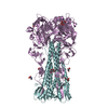
 Movie
Movie Controller


+ Open data

- Basic information
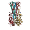
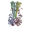
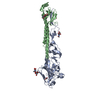
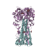
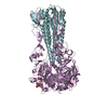
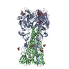
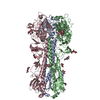
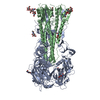
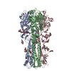
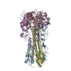
| Entry | Database: PDB / ID: 1rvz | |||||||||
|---|---|---|---|---|---|---|---|---|---|---|
| Title | 1934 H1 Hemagglutinin in complex with LSTC | |||||||||
 Components Components | (hemagglutinin) x 2 | |||||||||
 Keywords Keywords | VIRAL PROTEIN / Hemagglutinin / Influenza A Virus | |||||||||
| Function / homology |  Function and homology information Function and homology informationTransport of HA trimer, NA tetramer and M2 tetramer from the endoplasmic reticulum to the Golgi Apparatus / Assembly of Viral Components at the Budding Site / Influenza Infection / Fusion of the Influenza Virion to the Host Cell Endosome / Release / Budding / Packaging of Eight RNA Segments / Uncoating of the Influenza Virion / Entry of Influenza Virion into Host Cell via Endocytosis / Viral mRNA Translation ...Transport of HA trimer, NA tetramer and M2 tetramer from the endoplasmic reticulum to the Golgi Apparatus / Assembly of Viral Components at the Budding Site / Influenza Infection / Fusion of the Influenza Virion to the Host Cell Endosome / Release / Budding / Packaging of Eight RNA Segments / Uncoating of the Influenza Virion / Entry of Influenza Virion into Host Cell via Endocytosis / Viral mRNA Translation / viral budding from plasma membrane / Immunoregulatory interactions between a Lymphoid and a non-Lymphoid cell / clathrin-dependent endocytosis of virus by host cell / host cell surface receptor binding / fusion of virus membrane with host plasma membrane / fusion of virus membrane with host endosome membrane / viral envelope / virion attachment to host cell / host cell plasma membrane / virion membrane / extracellular region / membrane / plasma membrane Similarity search - Function | |||||||||
| Biological species |   Influenza A virus Influenza A virus | |||||||||
| Method |  X-RAY DIFFRACTION / SYNCHROTRON / MOLECULAR REPLACEMENT / Resolution: 2.25 Å X-RAY DIFFRACTION / SYNCHROTRON / MOLECULAR REPLACEMENT / Resolution: 2.25 Å | |||||||||
 Authors Authors | Gamblin, S.J. / Haire, L.F. / Russell, R.J. / Stevens, D.J. / Xiao, B. / Ha, Y. / Vasisht, N. / Steinhauer, D.A. / Daniels, R.S. / Elliot, A. ...Gamblin, S.J. / Haire, L.F. / Russell, R.J. / Stevens, D.J. / Xiao, B. / Ha, Y. / Vasisht, N. / Steinhauer, D.A. / Daniels, R.S. / Elliot, A. / Wiley, D.C. / Skehel, J.J. | |||||||||
 Citation Citation | Journal: Science / Year: 2004 Title: The structure and receptor binding properties of the 1918 influenza hemagglutinin. Authors: Gamblin, S.J. / Haire, L.F. / Russell, R.J. / Stevens, D.J. / Xiao, B. / Ha, Y. / Vasisht, N. / Steinhauer, D.A. / Daniels, R.S. / Elliot, A. / Wiley, D.C. / Skehel, J.J. | |||||||||
| History |
| |||||||||
| Remark 999 | SEQUENCE Author states that the sequence of this protein is correct and there is no matched ...SEQUENCE Author states that the sequence of this protein is correct and there is no matched sequence database available. Residue (E ASN 42 ) and residue (E GLY 44 ) are not linked, distance of C-N bond is 2.61. |
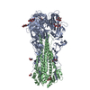
- Structure visualization
| Structure viewer | Molecule: MolmilJmol/JSmol |
|---|
- Downloads & links
-Download
| PDBx/mmCIF format | 1rvz.cif.gz | 622.9 KB | Display | PDBx/mmCIF format |
|---|---|---|---|---|
| PDB format | pdb1rvz.ent.gz | 510.8 KB | Display | PDB format |
| PDBx/mmJSON format | 1rvz.json.gz | Tree view | PDBx/mmJSON format | |
| Others |  Other downloads Other downloads |
-Validation report
| Arichive directory | https://data.pdbj.org/pub/pdb/validation_reports/rv/1rvzftp://data.pdbj.org/pub/pdb/validation_reports/rv/1rvz | HTTPS FTP |
|---|
-Related structure data
| Related structure data |  1ru7SC 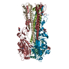 1ruyC 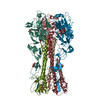 1ruzC 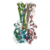 1rv0C 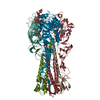 1rvtC 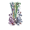 1rvxC S: Starting model for refinement C: citing same article ( |
|---|---|
| Similar structure data |


-Links
 PDBj
PDBj


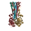
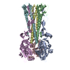
- Assembly
| Deposited unit | 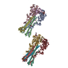
| ||||||||
|---|---|---|---|---|---|---|---|---|---|
| 1 | 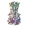
| ||||||||
| 2 | 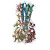
| ||||||||
| Unit cell |
|
-Components
| #1: Protein | Mass: 36733.402 Da / Num. of mol.: 6 / Source method: isolated from a natural source / Details: Bromelain digestion Source: (natural) Influenza A virus (A/Puerto Rico/8/34(H1N1))Genus: Influenzavirus A / Species: Influenza A virus / Strain: A-PUERTO RICO-8-34 / References: UniProt: Q82766, UniProt: P03452*PLUS #2: Protein | Mass: 18242.221 Da / Num. of mol.: 6 / Source method: isolated from a natural source / Details: Bromelain digestion Source: (natural) Influenza A virus (A/Puerto Rico/8/34(H1N1))Genus: Influenzavirus A / Species: Influenza A virus / Strain: A-PUERTO RICO-8-34 / References: UniProt: Q82766, UniProt: P03452*PLUS #3: Polysaccharide | N-acetyl-alpha-neuraminic acid-(2-6)-beta-D-galactopyranose-(1-4)-2-acetamido-2-deoxy-beta-D-glucopyranose / 6'-sialyl-N-acetyllactosamine 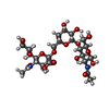  Source method: isolated from a genetically manipulated source Details: oligosaccharide / References: 6'-sialyl-N-acetyllactosamine #4: Sugar |   Type: D-saccharide, beta linking / Mass: 221.208 Da / Num. of mol.: 3 Type: D-saccharide, beta linking / Mass: 221.208 Da / Num. of mol.: 3Source method: isolated from a genetically manipulated source Formula: C8H15NO6 #5: Water | ChemComp-HOH / |  Mass: 18.015 Da / Num. of mol.: 2098 / Source method: isolated from a natural source / Formula: H2O Mass: 18.015 Da / Num. of mol.: 2098 / Source method: isolated from a natural source / Formula: H2OHas protein modification | Y | |
|---|
-Experimental details
-Experiment
| Experiment | Method: X-RAY DIFFRACTION / Number of used crystals: 1 |
|---|
- Sample preparation
| Crystal | Density Matthews: 3.73 Å3/Da / Density % sol: 67.01 % |
|---|---|
| Crystal grow | Temperature: 291 K / Method: vapor diffusion, hanging drop / pH: 7.5 Details: PEG3350, TrisHCl, KSCN, pH 7.5, VAPOR DIFFUSION, HANGING DROP, temperature 291K |
-Data collection
| Diffraction | Mean temperature: 100 K |
|---|---|
| Diffraction source | Source: SYNCHROTRON / Site: ESRF  / Beamline: ID14-1 / Wavelength: 0.933 / Beamline: ID14-1 / Wavelength: 0.933 |
| Detector | Type: ADSC QUANTUM 4 / Detector: CCD / Date: Sep 20, 2002 |
| Radiation | Protocol: SINGLE WAVELENGTH / Monochromatic (M) / Laue (L): M / Scattering type: x-ray |
| Radiation wavelength | Wavelength: 0.933 Å / Relative weight: 1 |
| Reflection | Resolution: 2.25→20 Å / Num. all: 229143 / Num. obs: 206016 / % possible obs: 89.9 % / Observed criterion σ(F): 0 / Observed criterion σ(I): 0 / Redundancy: 11.6 % / Rmerge(I) obs: 0.082 / Net I/σ(I): 14.6 |
| Reflection shell | Resolution: 2.25→2.33 Å / Rmerge(I) obs: 0.401 / Mean I/σ(I) obs: 2.9 / % possible all: 52.2 |
- Processing
| Software |
| ||||||||||||||||||||
|---|---|---|---|---|---|---|---|---|---|---|---|---|---|---|---|---|---|---|---|---|---|
| Refinement | Method to determine structure: MOLECULAR REPLACEMENT Starting model: PDB entry 1RU7 Resolution: 2.25→20 Å / σ(F): 0 / Stereochemistry target values: Engh & Huber
| ||||||||||||||||||||
| Refinement step | Cycle: LAST / Resolution: 2.25→20 Å
|
