 Movie
Movie Controller

[English] 日本語
 Yorodumi
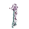
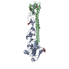
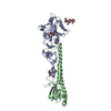
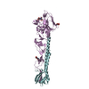
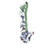
- PDB-1jsn: STRUCTURE OF AVIAN H5 HAEMAGGLUTININ COMPLEXED WITH LSTA RECEPTRO... -
+ Open data

- Basic information
| Entry | Database: PDB / ID: 1jsn | |||||||||
|---|---|---|---|---|---|---|---|---|---|---|
| Title | STRUCTURE OF AVIAN H5 HAEMAGGLUTININ COMPLEXED WITH LSTA RECEPTRO ANALOG | |||||||||
 Components Components | (HAEMAGGLUTININ ...) x 2 | |||||||||
 Keywords Keywords | VIRAL PROTEIN / influenza / receptor complex / fusion protein | |||||||||
| Function / homology |  Function and homology information Function and homology informationviral budding from plasma membrane / clathrin-dependent endocytosis of virus by host cell / host cell surface receptor binding / fusion of virus membrane with host plasma membrane / fusion of virus membrane with host endosome membrane / viral envelope / virion attachment to host cell / host cell plasma membrane / virion membrane / membrane Similarity search - Function | |||||||||
| Biological species |   Influenza A virus Influenza A virus | |||||||||
| Method |  X-RAY DIFFRACTION / SYNCHROTRON / MOLECULAR REPLACEMENT / Resolution: 2.4 Å X-RAY DIFFRACTION / SYNCHROTRON / MOLECULAR REPLACEMENT / Resolution: 2.4 Å | |||||||||
 Authors Authors | Ha, Y. / Stevens, D.J. / Skehel, J.J. / Wiley, D.C. | |||||||||
 Citation Citation | Journal: Proc.Natl.Acad.Sci.USA / Year: 2001 Title: X-ray structures of H5 avian and H9 swine influenza virus hemagglutinins bound to avian and human receptor analogs. Authors: Ha, Y. / Stevens, D.J. / Skehel, J.J. / Wiley, D.C. | |||||||||
| History |
|
- Structure visualization
| Structure viewer | Molecule: MolmilJmol/JSmol |
|---|
- Downloads & links
-Download
| PDBx/mmCIF format | 1jsn.cif.gz | 116.1 KB | Display | PDBx/mmCIF format |
|---|---|---|---|---|
| PDB format | pdb1jsn.ent.gz | 89.6 KB | Display | PDB format |
| PDBx/mmJSON format | 1jsn.json.gz | Tree view | PDBx/mmJSON format | |
| Others |  Other downloads Other downloads |
-Validation report
| Arichive directory | https://data.pdbj.org/pub/pdb/validation_reports/js/1jsnftp://data.pdbj.org/pub/pdb/validation_reports/js/1jsn | HTTPS FTP |
|---|
-Related structure data


-Links
 PDBj
PDBj


- Assembly
| Deposited unit | 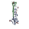
| ||||||||
|---|---|---|---|---|---|---|---|---|---|
| 1 |
| ||||||||
| Unit cell |
|
-Components
-HAEMAGGLUTININ ... , 2 types, 2 molecules AB
| #1: Protein | Mass: 36573.207 Da / Num. of mol.: 1 / Fragment: Residues 1-325 / Source method: isolated from a natural source / Details: AUTO CLEAVED FRAGMENT OF N-TERMINUS / Source: (natural) Influenza A virus / Genus: Influenzavirus A / Strain: A/Duck/Singapore/3/97 / References: UniProt: A5Z226 |
|---|---|
| #2: Protein | Mass: 20200.232 Da / Num. of mol.: 1 / Fragment: Residues 1-176 / Source method: isolated from a natural source / Details: AUTO CLEAVED FRAGMENT OF C-TERMINUS / Source: (natural) Influenza A virus / Genus: Influenzavirus A / Strain: A/Duck/Singapore/3/97 / References: UniProt: A5Z226*PLUS |
-Sugars , 3 types, 6 molecules 
| #3: Polysaccharide | Source method: isolated from a genetically manipulated source #4: Polysaccharide | N-acetyl-alpha-neuraminic acid-(2-3)-beta-D-galactopyranose-(1-3)-2-acetamido-2-deoxy-beta-D-glucopyranose | Source method: isolated from a genetically manipulated source #5: Sugar |  Type: D-saccharide, beta linking / Mass: 221.208 Da / Num. of mol.: 3 Type: D-saccharide, beta linking / Mass: 221.208 Da / Num. of mol.: 3Source method: isolated from a genetically manipulated source Formula: C8H15NO6 |
|---|
-Non-polymers , 1 types, 216 molecules 
| #6: Water | ChemComp-HOH / Mass: 18.015 Da / Num. of mol.: 216 / Source method: isolated from a natural source / Formula: H2O |
|---|
-Details
| Has protein modification | Y |
|---|
-Experimental details
-Experiment
| Experiment | Method: X-RAY DIFFRACTION / Number of used crystals: 1 |
|---|
- Sample preparation
| Crystal | Density Matthews: 3.73 Å3/Da / Density % sol: 67.02 % |
|---|---|
| Crystal grow | Temperature: 298 K / Method: vapor diffusion, hanging drop / pH: 7.5 Details: mme-peg 2000, nicl2, hepes, pH 7.5, VAPOR DIFFUSION, HANGING DROP, temperature 298K |
| Crystal grow | *PLUS Method: unknown |
-Data collection
| Diffraction | Mean temperature: 100 K |
|---|---|
| Diffraction source | Source: SYNCHROTRON / Site: APS  / Beamline: 14-BM-C / Wavelength: 1 Å / Beamline: 14-BM-C / Wavelength: 1 Å |
| Detector | Type: ADSC QUANTUM 4 / Detector: CCD / Date: Dec 31, 1999 |
| Radiation | Monochromator: graphite / Protocol: SINGLE WAVELENGTH / Monochromatic (M) / Laue (L): M / Scattering type: x-ray |
| Radiation wavelength | Wavelength: 1 Å / Relative weight: 1 |
| Reflection | Resolution: 2.4→40 Å / Num. all: 33434 / Num. obs: 27009 / % possible obs: 80.8 % / Observed criterion σ(F): 1 / Observed criterion σ(I): 1 |
| Reflection shell | Resolution: 2.4→40 Å / % possible all: 81 |
| Reflection | *PLUS Lowest resolution: 40 Å / Num. obs: 30623 / % possible obs: 92.7 % / Rmerge(I) obs: 0.106 |
| Reflection shell | *PLUS % possible obs: 94 % / Rmerge(I) obs: 0.709 |
- Processing
| Software |
| ||||||||||||||||||||||||||||||
|---|---|---|---|---|---|---|---|---|---|---|---|---|---|---|---|---|---|---|---|---|---|---|---|---|---|---|---|---|---|---|---|
| Refinement | Method to determine structure: MOLECULAR REPLACEMENT / Resolution: 2.4→40 Å / σ(F): 0 / Stereochemistry target values: Engh & Huber
| ||||||||||||||||||||||||||||||
| Refinement step | Cycle: LAST / Resolution: 2.4→40 Å
| ||||||||||||||||||||||||||||||
| Software | *PLUS Name: CNS / Classification: refinement | ||||||||||||||||||||||||||||||
| Refinement | *PLUS Highest resolution: 2.4 Å / Lowest resolution: 40 Å / σ(F): 0 / Rfactor obs: 0.24 / Rfactor Rwork: 0.24 | ||||||||||||||||||||||||||||||
| Solvent computation | *PLUS | ||||||||||||||||||||||||||||||
| Displacement parameters | *PLUS | ||||||||||||||||||||||||||||||
| Refine LS restraints | *PLUS
| ||||||||||||||||||||||||||||||
| LS refinement shell | *PLUS Rfactor Rfree: 0.634 / Rfactor obs: 0.588 |
