 Movie
Movie Controller

[English] 日本語
 Yorodumi
- PDB-5vjk: Crystal structure of H7 hemagglutinin mutant (V186K, K193T, G228S... -
+ Open data

- Basic information
| Entry | Database: PDB / ID: 5vjk | |||||||||
|---|---|---|---|---|---|---|---|---|---|---|
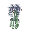
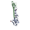
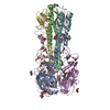
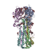
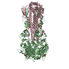
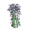
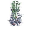
| Title | Crystal structure of H7 hemagglutinin mutant (V186K, K193T, G228S) from the influenza virus A/Shanghai/2/2013 (H7N9) | |||||||||
 Components Components |
| |||||||||
 Keywords Keywords | VIRAL PROTEIN / influenza virus / hemagglutinin / mutant / receptor specificity | |||||||||
| Function / homology |  Function and homology information Function and homology informationviral budding from plasma membrane / clathrin-dependent endocytosis of virus by host cell / host cell surface receptor binding / fusion of virus membrane with host plasma membrane / fusion of virus membrane with host endosome membrane / viral envelope / virion attachment to host cell / host cell plasma membrane / virion membrane / membrane / metal ion binding Similarity search - Function | |||||||||
| Biological species |   Influenza A virus Influenza A virus | |||||||||
| Method |  X-RAY DIFFRACTION / SYNCHROTRON / MOLECULAR REPLACEMENT / Resolution: 2.593 Å X-RAY DIFFRACTION / SYNCHROTRON / MOLECULAR REPLACEMENT / Resolution: 2.593 Å | |||||||||
 Authors Authors | Zhu, X. / Wilson, I.A. | |||||||||
 Citation Citation | Journal: PLoS Pathog. / Year: 2017 Title: Three mutations switch H7N9 influenza to human-type receptor specificity. Authors: de Vries, R.P. / Peng, W. / Grant, O.C. / Thompson, A.J. / Zhu, X. / Bouwman, K.M. / de la Pena, A.T.T. / van Breemen, M.J. / Ambepitiya Wickramasinghe, I.N. / de Haan, C.A.M. / Yu, W. / ...Authors: de Vries, R.P. / Peng, W. / Grant, O.C. / Thompson, A.J. / Zhu, X. / Bouwman, K.M. / de la Pena, A.T.T. / van Breemen, M.J. / Ambepitiya Wickramasinghe, I.N. / de Haan, C.A.M. / Yu, W. / McBride, R. / Sanders, R.W. / Woods, R.J. / Verheije, M.H. / Wilson, I.A. / Paulson, J.C. | |||||||||
| History |
|
- Structure visualization
| Structure viewer | Molecule: MolmilJmol/JSmol |
|---|
- Downloads & links
-Download
| PDBx/mmCIF format | 5vjk.cif.gz | 210.3 KB | Display | PDBx/mmCIF format |
|---|---|---|---|---|
| PDB format | pdb5vjk.ent.gz | 167.4 KB | Display | PDB format |
| PDBx/mmJSON format | 5vjk.json.gz | Tree view | PDBx/mmJSON format | |
| Others |  Other downloads Other downloads |
-Validation report
| Arichive directory | https://data.pdbj.org/pub/pdb/validation_reports/vj/5vjkftp://data.pdbj.org/pub/pdb/validation_reports/vj/5vjk | HTTPS FTP |
|---|
-Related structure data
| Related structure data | 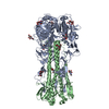 5vjlC 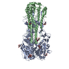 5vjmC 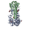 4n5jS C: citing same article ( S: Starting model for refinement |
|---|---|
| Similar structure data |


-Links
 PDBj
PDBj


- Assembly
| Deposited unit | 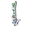
| ||||||||
|---|---|---|---|---|---|---|---|---|---|
| 1 | 
| ||||||||
| Unit cell |
| ||||||||
| Components on special symmetry positions |
|
-Components
| #1: Protein | Mass: 35365.891 Da / Num. of mol.: 1 / Mutation: V186K, K193T, G228S Source method: isolated from a genetically manipulated source Source: (gene. exp.) Influenza A virus / Strain: A/Shanghai/02/2013(H7N9) / Gene: HA / Production host:  Trichoplusia ni (cabbage looper) / References: UniProt: R4NN21 Trichoplusia ni (cabbage looper) / References: UniProt: R4NN21 | ||||
|---|---|---|---|---|---|
| #2: Protein | Mass: 21081.207 Da / Num. of mol.: 1 Source method: isolated from a genetically manipulated source Source: (gene. exp.) Influenza A virus / Strain: A/Shanghai/02/2013(H7N9) / Gene: HA / Production host: Trichoplusia ni (cabbage looper) / References: UniProt: R4NN21 | ||||
| #3: Polysaccharide | beta-D-mannopyranose-(1-4)-2-acetamido-2-deoxy-beta-D-glucopyranose-(1-4)-2-acetamido-2-deoxy-beta- ...beta-D-mannopyranose-(1-4)-2-acetamido-2-deoxy-beta-D-glucopyranose-(1-4)-2-acetamido-2-deoxy-beta-D-glucopyranose Source method: isolated from a genetically manipulated source | ||||
| #4: Sugar |   Type: D-saccharide, beta linking / Mass: 221.208 Da / Num. of mol.: 2 Type: D-saccharide, beta linking / Mass: 221.208 Da / Num. of mol.: 2Source method: isolated from a genetically manipulated source Formula: C8H15NO6 #5: Water | ChemComp-HOH / |  Mass: 18.015 Da / Num. of mol.: 71 / Source method: isolated from a natural source / Formula: H2O Mass: 18.015 Da / Num. of mol.: 71 / Source method: isolated from a natural source / Formula: H2OHas protein modification | Y | |
-Experimental details
-Experiment
| Experiment | Method: X-RAY DIFFRACTION / Number of used crystals: 1 |
|---|
- Sample preparation
| Crystal | Density Matthews: 3.35 Å3/Da / Density % sol: 63.28 % |
|---|---|
| Crystal grow | Temperature: 295 K / Method: vapor diffusion, sitting drop / pH: 8 Details: 0.2 M tri-potassium citrate, 5% (v/v) ethylene glycol and 22% (w/v) PEG3350 |
-Data collection
| Diffraction | Mean temperature: 100 K |
|---|---|
| Diffraction source | Source: SYNCHROTRON / Site: APS  / Beamline: 21-ID-D / Wavelength: 1.033 Å / Beamline: 21-ID-D / Wavelength: 1.033 Å |
| Detector | Type: DECTRIS PILATUS3 6M / Detector: PIXEL / Date: Mar 23, 2014 |
| Radiation | Protocol: SINGLE WAVELENGTH / Monochromatic (M) / Laue (L): M / Scattering type: x-ray |
| Radiation wavelength | Wavelength: 1.033 Å / Relative weight: 1 |
| Reflection | Resolution: 2.593→50 Å / Num. obs: 23692 / % possible obs: 99.3 % / Redundancy: 4 % / CC1/2: 0.998 / Rpim(I) all: 0.05 / Rsym value: 0.09 / Net I/σ(I): 16.4 |
| Reflection shell | Resolution: 2.593→2.64 Å / Redundancy: 2.8 % / Num. unique obs: 1102 / CC1/2: 0.548 / Rpim(I) all: 0.44 / Rsym value: 0.66 / % possible all: 93.4 |
- Processing
| Software |
| ||||||||||||||||||||||||||||||||||||||||||||||||||||||||||||||||||||||
|---|---|---|---|---|---|---|---|---|---|---|---|---|---|---|---|---|---|---|---|---|---|---|---|---|---|---|---|---|---|---|---|---|---|---|---|---|---|---|---|---|---|---|---|---|---|---|---|---|---|---|---|---|---|---|---|---|---|---|---|---|---|---|---|---|---|---|---|---|---|---|---|
| Refinement | Method to determine structure: MOLECULAR REPLACEMENT Starting model: 4N5J Resolution: 2.593→49.772 Å / SU ML: 0.4 / Cross valid method: THROUGHOUT / σ(F): 1.35 / Phase error: 25.82 / Stereochemistry target values: ML
| ||||||||||||||||||||||||||||||||||||||||||||||||||||||||||||||||||||||
| Solvent computation | Shrinkage radii: 0.9 Å / VDW probe radii: 1.11 Å / Solvent model: FLAT BULK SOLVENT MODEL | ||||||||||||||||||||||||||||||||||||||||||||||||||||||||||||||||||||||
| Refinement step | Cycle: LAST / Resolution: 2.593→49.772 Å
| ||||||||||||||||||||||||||||||||||||||||||||||||||||||||||||||||||||||
| Refine LS restraints |
| ||||||||||||||||||||||||||||||||||||||||||||||||||||||||||||||||||||||
| LS refinement shell |
| ||||||||||||||||||||||||||||||||||||||||||||||||||||||||||||||||||||||
| Refinement TLS params. | Method: refined / Origin x: -52.8954 Å / Origin y: 17.8726 Å / Origin z: 7.9076 Å
| ||||||||||||||||||||||||||||||||||||||||||||||||||||||||||||||||||||||
| Refinement TLS group | Selection details: all |
