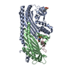
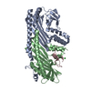
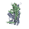
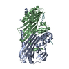
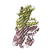
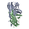
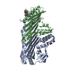
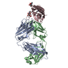
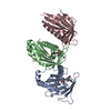
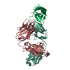
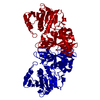
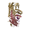
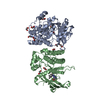
登録情報 データベース : PDB / ID : 3aaaタイトル Crystal Structure of Actin capping protein in complex with V-1 F-actin-capping protein subunit alpha-1 F-actin-capping protein subunit beta isoforms 1 and 2 Myotrophin キーワード / / / / / / / 機能・相同性 分子機能 ドメイン・相同性 構成要素
/ / / / / / / / / / / / / / / / / / / / / / / / / / / / / / / / / / / / / / / / / / / / / / / / / / / / / / / / / / / / / / / / / / / / / / / / / / / / / / / / / / / / / / / / / / / / / / / / 生物種 Gallus gallus (ニワトリ)Homo sapiens (ヒト)手法 / / / 解像度 : 2.2 Å データ登録者 Takeda, S. / Minakata, S. / Narita, A. / Kitazawa, M. / Yamakuni, T. / Maeda, Y. / Nitanai, Y. ジャーナル : Plos Biol. / 年 : 2010タイトル : Two distinct mechanisms for actin capping protein regulation--steric and allosteric inhibition著者 : Takeda, S. / Minakata, S. / Koike, R. / Kawahata, I. / Narita, A. / Kitazawa, M. / Ota, M. / Yamakuni, T. / Maeda, Y. / Nitanai, Y. 履歴 登録 2009年11月12日 登録サイト / 処理サイト 改定 1.0 2010年8月4日 Provider / タイプ 改定 1.1 2011年7月13日 Group 改定 1.2 2023年11月1日 Group Data collection / Database references ... Data collection / Database references / Derived calculations / Refinement description カテゴリ chem_comp_atom / chem_comp_bond ... chem_comp_atom / chem_comp_bond / database_2 / pdbx_initial_refinement_model / struct_ref_seq_dif / struct_site Item _database_2.pdbx_DOI / _database_2.pdbx_database_accession ... _database_2.pdbx_DOI / _database_2.pdbx_database_accession / _struct_ref_seq_dif.details / _struct_site.pdbx_auth_asym_id / _struct_site.pdbx_auth_comp_id / _struct_site.pdbx_auth_seq_id
すべて表示 表示を減らす
 ムービー
ムービー コントローラー


 データを開く
 基本情報
基本情報 要素
要素 キーワード
キーワード 機能・相同性情報
 Homo sapiens (ヒト)
 X線回折 /
  データ登録者
 引用
引用 構造の表示
構造の表示 ダウンロードとリンク
 その他のダウンロード
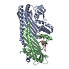


 PDBj
PDBj
 集合体
集合体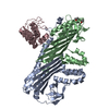


 分子量: 60.095 Da / 分子数: 2 / 由来タイプ: 合成 / 式: C3H8O
 分子量: 18.015 Da / 分子数: 354 / 由来タイプ: 天然 / 式: H2O
 試料調製
試料調製 / ビームライン: BL26B1 / 波長: 1 Å
 解析
解析