 Movie
Movie Controller

[English] 日本語
 Yorodumi
- PDB-3aa0: Crystal structure of Actin Capping Protein in complex with the Cp... -
+ Open data

- Basic information
| Entry | Database: PDB / ID: 3aa0 | ||||||
|---|---|---|---|---|---|---|---|
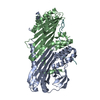
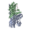
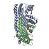
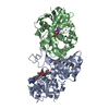
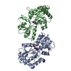
| Title | Crystal structure of Actin Capping Protein in complex with the Cp-binding motif derived from CARMIL | ||||||
 Components Components |
| ||||||
 Keywords Keywords | PROTEIN BINDING / ACTIN CAPPING PROTEIN / BARBED END REGULATION / CARMIL FAMILY PROTEIN / CONFORMATIONAL CHANGE / CELL MOTILITY / Actin capping / Actin-binding / Cytoskeleton / Isopeptide bond / Leucine-rich repeat | ||||||
| Function / homology |  Function and homology information Function and homology informationbarbed-end actin filament uncapping / positive regulation of lamellipodium organization / negative regulation of barbed-end actin filament capping / Advanced glycosylation endproduct receptor signaling / : / RHOF GTPase cycle / HSP90 chaperone cycle for steroid hormone receptors (SHR) in the presence of ligand / COPI-independent Golgi-to-ER retrograde traffic / macropinosome / Factors involved in megakaryocyte development and platelet production ...barbed-end actin filament uncapping / positive regulation of lamellipodium organization / negative regulation of barbed-end actin filament capping / Advanced glycosylation endproduct receptor signaling / : / RHOF GTPase cycle / HSP90 chaperone cycle for steroid hormone receptors (SHR) in the presence of ligand / COPI-independent Golgi-to-ER retrograde traffic / macropinosome / Factors involved in megakaryocyte development and platelet production / COPI-mediated anterograde transport / negative regulation of filopodium assembly / actin filament network formation / F-actin capping protein complex / WASH complex / macropinocytosis / regulation of Arp2/3 complex-mediated actin nucleation / Factors involved in megakaryocyte development and platelet production / urate metabolic process / cell junction assembly / ruffle organization / barbed-end actin filament capping / actin polymerization or depolymerization / regulation of lamellipodium assembly / intermediate filament cytoskeleton / regulation of cell morphogenesis / lamellipodium assembly / filamentous actin / positive regulation of actin filament polymerization / cell leading edge / cortical cytoskeleton / establishment or maintenance of cell polarity / brush border / sperm head-tail coupling apparatus / positive regulation of stress fiber assembly / cytoskeleton organization / positive regulation of substrate adhesion-dependent cell spreading / Schaffer collateral - CA1 synapse / cell morphogenesis / Z disc / actin filament binding / cell migration / lamellipodium / actin cytoskeleton organization / nuclear speck / postsynaptic density / positive regulation of cell migration / protein-containing complex binding / membrane / nucleus / plasma membrane / cytoplasm / cytosol Similarity search - Function | ||||||
| Biological species |   | ||||||
| Method |  X-RAY DIFFRACTION / SYNCHROTRON / MOLECULAR REPLACEMENT / Resolution: 1.7 Å X-RAY DIFFRACTION / SYNCHROTRON / MOLECULAR REPLACEMENT / Resolution: 1.7 Å | ||||||
 Authors Authors | Takeda, S. / Minakata, S. / Narita, A. / Kitazawa, M. / Yamakuni, T. / Maeda, Y. / Nitanai, Y. | ||||||
 Citation Citation | Journal: Plos Biol. / Year: 2010 Title: Two distinct mechanisms for actin capping protein regulation--steric and allosteric inhibition Authors: Takeda, S. / Minakata, S. / Koike, R. / Kawahata, I. / Narita, A. / Kitazawa, M. / Ota, M. / Yamakuni, T. / Maeda, Y. / Nitanai, Y. | ||||||
| History |
|
- Structure visualization
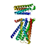
| Structure viewer | Molecule: MolmilJmol/JSmol |
|---|
- Downloads & links
-Download
| PDBx/mmCIF format | 3aa0.cif.gz | 132.6 KB | Display | PDBx/mmCIF format |
|---|---|---|---|---|
| PDB format | pdb3aa0.ent.gz | 101.6 KB | Display | PDB format |
| PDBx/mmJSON format | 3aa0.json.gz | Tree view | PDBx/mmJSON format | |
| Others |  Other downloads Other downloads |
-Validation report
| Arichive directory | https://data.pdbj.org/pub/pdb/validation_reports/aa/3aa0ftp://data.pdbj.org/pub/pdb/validation_reports/aa/3aa0 | HTTPS FTP |
|---|
-Related structure data
| Related structure data |  3aa1C 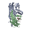 3aa6C 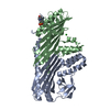 3aa7C 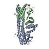 3aaaC 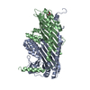 1iznS S: Starting model for refinement C: citing same article ( |
|---|---|
| Similar structure data |
-Links
 PDBj
PDBj


- Assembly
| Deposited unit | 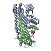
| ||||||||
|---|---|---|---|---|---|---|---|---|---|
| 1 |
| ||||||||
| Unit cell |
| ||||||||
| Details | THE ORIGOMETRIC STATE OF THE ACTIN CAPPING PROTEIN IS A HETERO DIMER COMPOSE OF SUBUNIT A (CHAIN A) AND SUBUNIT B (CHAIN B) IN VIVO AND IN VITRO. |
-Components
| #1: Protein | Mass: 33001.789 Da / Num. of mol.: 1 Source method: isolated from a genetically manipulated source Source: (gene. exp.)  |
|---|---|
| #2: Protein | Mass: 27473.070 Da / Num. of mol.: 1 / Mutation: residues 244-277 deletion mutation Source method: isolated from a genetically manipulated source Details: BETA TENTACLE DELETION / Source: (gene. exp.) |
| #3: Protein/peptide | Mass: 2613.070 Da / Num. of mol.: 1 / Fragment: CP-BINDING MOTIF, recidues 985-1005 / Source method: obtained synthetically / Details: THE PEPTIDE WAS CHEMICALLY SYNTHESIZED. / Source: (synth.) |
| #4: Chemical | ChemComp-CO3 /   Mass: 60.009 Da / Num. of mol.: 1 / Source method: obtained synthetically / Formula: CO3 Mass: 60.009 Da / Num. of mol.: 1 / Source method: obtained synthetically / Formula: CO3 |
| #5: Water | ChemComp-HOH /  Mass: 18.015 Da / Num. of mol.: 438 / Source method: isolated from a natural source / Formula: H2O Mass: 18.015 Da / Num. of mol.: 438 / Source method: isolated from a natural source / Formula: H2O |
-Experimental details
-Experiment
| Experiment | Method: X-RAY DIFFRACTION / Number of used crystals: 1 |
|---|
- Sample preparation
| Crystal | Density Matthews: 2.02 Å3/Da / Density % sol: 39.18 % |
|---|---|
| Crystal grow | Temperature: 293 K / Method: vapor diffusion, hanging drop / pH: 6 Details: 18% PEG 400, 40MM BACL2, 100MM MES-NAOH, PH 6.0, VAPOR DIFFUSION, HANGING DROP, temperature 293.0K |
-Data collection
| Diffraction | Mean temperature: 100 K |
|---|---|
| Diffraction source | Source: SYNCHROTRON / Site: SPring-8  / Beamline: BL26B1 / Wavelength: 1 Å / Beamline: BL26B1 / Wavelength: 1 Å |
| Detector | Type: RIGAKU JUPITER 210 / Detector: CCD / Date: Feb 12, 2009 / Details: mirrors |
| Radiation | Monochromator: SI / Protocol: SINGLE WAVELENGTH / Monochromatic (M) / Laue (L): M / Scattering type: x-ray |
| Radiation wavelength | Wavelength: 1 Å / Relative weight: 1 |
| Reflection | Resolution: 1.7→50 Å / Num. obs: 55697 / % possible obs: 97.7 % / Redundancy: 6.1 % / Biso Wilson estimate: 33.5 Å2 / Rmerge(I) obs: 0.08 / Net I/σ(I): 18.32 |
| Reflection shell | Resolution: 1.7→1.76 Å / Redundancy: 2.5 % / Rmerge(I) obs: 0.348 / Mean I/σ(I) obs: 2.03 / Num. unique all: 4405 / % possible all: 78.7 |
- Processing
| Software |
| |||||||||||||||||||||||||||||||||||||||||||||||||||||||||||||||||
|---|---|---|---|---|---|---|---|---|---|---|---|---|---|---|---|---|---|---|---|---|---|---|---|---|---|---|---|---|---|---|---|---|---|---|---|---|---|---|---|---|---|---|---|---|---|---|---|---|---|---|---|---|---|---|---|---|---|---|---|---|---|---|---|---|---|---|
| Refinement | Method to determine structure: MOLECULAR REPLACEMENT Starting model: PDB ENTRY 1IZN Resolution: 1.7→42.81 Å / Cor.coef. Fo:Fc: 0.957 / Cor.coef. Fo:Fc free: 0.929 / SU B: 2.382 / SU ML: 0.079 / Cross valid method: THROUGHOUT / ESU R: 0.127 / ESU R Free: 0.126 / Stereochemistry target values: MAXIMUM LIKELIHOOD / Details: HYDROGENS HAVE BEEN ADDED IN THE RIDING POSITIONS
| |||||||||||||||||||||||||||||||||||||||||||||||||||||||||||||||||
| Solvent computation | Ion probe radii: 0.8 Å / Shrinkage radii: 0.8 Å / VDW probe radii: 1.4 Å / Solvent model: MASK | |||||||||||||||||||||||||||||||||||||||||||||||||||||||||||||||||
| Displacement parameters | Biso mean: 26.033 Å2
| |||||||||||||||||||||||||||||||||||||||||||||||||||||||||||||||||
| Refine analyze | Luzzati coordinate error obs: 0.189 Å | |||||||||||||||||||||||||||||||||||||||||||||||||||||||||||||||||
| Refinement step | Cycle: LAST / Resolution: 1.7→42.81 Å
| |||||||||||||||||||||||||||||||||||||||||||||||||||||||||||||||||
| Refine LS restraints |
| |||||||||||||||||||||||||||||||||||||||||||||||||||||||||||||||||
| LS refinement shell | Resolution: 1.701→1.745 Å / Total num. of bins used: 20
|
