 Movie
Movie Controller


+ Open data

- Basic information
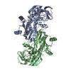
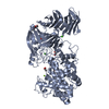
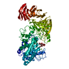
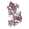
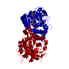
| Entry | Database: PDB / ID: 1izn | ||||||
|---|---|---|---|---|---|---|---|
| Title | Crystal Structure of Actin Filament Capping Protein CapZ | ||||||
 Components Components |
| ||||||
 Keywords Keywords | PROTEIN BINDING / HETERODIMER / CAPPING PROTEIN / ACTIN FILAMENT BARBED END CAPPING | ||||||
| Function / homology |  Function and homology information Function and homology informationAdvanced glycosylation endproduct receptor signaling / negative regulation of filopodium assembly / F-actin capping protein complex / WASH complex / cell junction assembly / barbed-end actin filament capping / regulation of lamellipodium assembly / sperm head-tail coupling apparatus / cortical cytoskeleton / brush border ...Advanced glycosylation endproduct receptor signaling / negative regulation of filopodium assembly / F-actin capping protein complex / WASH complex / cell junction assembly / barbed-end actin filament capping / regulation of lamellipodium assembly / sperm head-tail coupling apparatus / cortical cytoskeleton / brush border / cell morphogenesis / Schaffer collateral - CA1 synapse / Z disc / actin filament binding / lamellipodium / actin cytoskeleton organization / postsynaptic density / membrane / plasma membrane / cytosol Similarity search - Function | ||||||
| Biological species |  | ||||||
| Method |  X-RAY DIFFRACTION / SYNCHROTRON / MAD / Resolution: 2.1 Å X-RAY DIFFRACTION / SYNCHROTRON / MAD / Resolution: 2.1 Å | ||||||
 Authors Authors | Yamashita, A. / Maeda, K. / Maeda, Y. | ||||||
 Citation Citation | Journal: EMBO J. / Year: 2003 Title: Crystal structure of CapZ: structural basis for actin filament barbed end capping Authors: Yamashita, A. / Maeda, K. / Maeda, Y. | ||||||
| History |
|
- Structure visualization
| Structure viewer | Molecule: MolmilJmol/JSmol |
|---|
- Downloads & links
-Download
| PDBx/mmCIF format | 1izn.cif.gz | 235 KB | Display | PDBx/mmCIF format |
|---|---|---|---|---|
| PDB format | pdb1izn.ent.gz | 189.4 KB | Display | PDB format |
| PDBx/mmJSON format | 1izn.json.gz | Tree view | PDBx/mmJSON format | |
| Others |  Other downloads Other downloads |
-Validation report
| Arichive directory | https://data.pdbj.org/pub/pdb/validation_reports/iz/1iznftp://data.pdbj.org/pub/pdb/validation_reports/iz/1izn | HTTPS FTP |
|---|
-Related structure data
| Similar structure data |
|---|


-Links
 PDBj
PDBj- Assembly
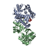
| Deposited unit | 
| ||||||||
|---|---|---|---|---|---|---|---|---|---|
| 1 | 
| ||||||||
| 2 | 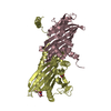
| ||||||||
| Unit cell |
| ||||||||
| Details | Chains A and B, C and D are biological heterodimer assemblies respectively. |
-Components
| #1: Protein | Mass: 33001.789 Da / Num. of mol.: 2 Source method: isolated from a genetically manipulated source Source: (gene. exp.)  #2: Protein | Mass: 31403.449 Da / Num. of mol.: 2 Source method: isolated from a genetically manipulated source Source: (gene. exp.) Description: The cDNAs encoding alpha-1 and beta-1 subunits were cloned in a single vector Plasmid: pET3d / Production host: #3: Chemical | ChemComp-NO3 /   Mass: 62.005 Da / Num. of mol.: 4 / Source method: obtained synthetically / Formula: NO3 Mass: 62.005 Da / Num. of mol.: 4 / Source method: obtained synthetically / Formula: NO3#4: Water | ChemComp-HOH / |  Mass: 18.015 Da / Num. of mol.: 513 / Source method: isolated from a natural source / Formula: H2O Mass: 18.015 Da / Num. of mol.: 513 / Source method: isolated from a natural source / Formula: H2O |
|---|
-Experimental details
-Experiment
| Experiment | Method: X-RAY DIFFRACTION / Number of used crystals: 1 |
|---|
- Sample preparation
| Crystal | Density Matthews: 2.01 Å3/Da / Density % sol: 38.8 % | ||||||||||||||||||||||||||||||||||||||||||||||||||||||||
|---|---|---|---|---|---|---|---|---|---|---|---|---|---|---|---|---|---|---|---|---|---|---|---|---|---|---|---|---|---|---|---|---|---|---|---|---|---|---|---|---|---|---|---|---|---|---|---|---|---|---|---|---|---|---|---|---|---|
| Crystal grow | Temperature: 293 K / Method: vapor diffusion, hanging drop / pH: 5 Details: PEG3350, magnesium nitrate, MES-NAOH, Jeffamine M-600, pH 5.0, VAPOR DIFFUSION, HANGING DROP, temperature 293.0K | ||||||||||||||||||||||||||||||||||||||||||||||||||||||||
| Crystal grow | *PLUS Temperature: 20 ℃ / pH: 8 | ||||||||||||||||||||||||||||||||||||||||||||||||||||||||
| Components of the solutions | *PLUS
|
-Data collection
| Diffraction | Mean temperature: 90 K |
|---|---|
| Diffraction source | Source: SYNCHROTRON / Site: SPring-8  / Beamline: BL45PX / Wavelength: 1.02 Å / Beamline: BL45PX / Wavelength: 1.02 Å |
| Detector | Type: RIGAKU RAXIS V / Detector: IMAGE PLATE / Date: Apr 24, 2002 / Details: CYLINDRICAL BEND MIRROR |
| Radiation | Monochromator: DIAMOND / Protocol: SINGLE WAVELENGTH / Monochromatic (M) / Laue (L): M / Scattering type: x-ray |
| Radiation wavelength | Wavelength: 1.02 Å / Relative weight: 1 |
| Reflection | Resolution: 2.1→50 Å / Num. all: 61111 / Num. obs: 61088 / % possible obs: 100 % / Observed criterion σ(I): -3 / Redundancy: 3.75 % / Biso Wilson estimate: 21.5 Å2 / Rmerge(I) obs: 0.051 / Net I/σ(I): 26 |
| Reflection shell | Resolution: 2.1→2.18 Å / Rmerge(I) obs: 0.265 / % possible all: 100 |
| Reflection | *PLUS Num. measured all: 228951 |
| Reflection shell | *PLUS % possible obs: 100 % |
- Processing
| Software |
| ||||||||||||||||||||||||||||||||||||
|---|---|---|---|---|---|---|---|---|---|---|---|---|---|---|---|---|---|---|---|---|---|---|---|---|---|---|---|---|---|---|---|---|---|---|---|---|---|
| Refinement | Method to determine structure: MAD / Resolution: 2.1→50 Å / Data cutoff high rms absF: 10000 / Cross valid method: THROUGHOUT / σ(F): 0 / Stereochemistry target values: Engh & Huber
| ||||||||||||||||||||||||||||||||||||
| Solvent computation | Solvent model: FLAT MODEL / Bsol: 49.24 Å2 / ksol: 0.34 e/Å3 | ||||||||||||||||||||||||||||||||||||
| Displacement parameters | Biso mean: 43.7 Å2
| ||||||||||||||||||||||||||||||||||||
| Refine analyze |
| ||||||||||||||||||||||||||||||||||||
| Refinement step | Cycle: LAST / Resolution: 2.1→50 Å
| ||||||||||||||||||||||||||||||||||||
| Refine LS restraints |
| ||||||||||||||||||||||||||||||||||||
| LS refinement shell | Resolution: 2.1→2.18 Å / Total num. of bins used: 10
| ||||||||||||||||||||||||||||||||||||
| Xplor file |
| ||||||||||||||||||||||||||||||||||||
| Refinement | *PLUS % reflection Rfree: 5 % | ||||||||||||||||||||||||||||||||||||
| Solvent computation | *PLUS | ||||||||||||||||||||||||||||||||||||
| Displacement parameters | *PLUS | ||||||||||||||||||||||||||||||||||||
| Refine LS restraints | *PLUS
|
