- PDB-4gvo: Putative L-Cystine ABC transporter from Listeria monocytogenes -
+
Open data
ID or keywords:
Loading...
-
Basic information
Entry
Database: PDB / ID: 4gvo
Title
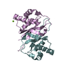
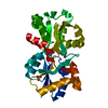
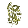
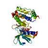
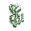
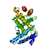
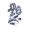
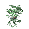
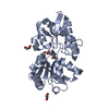
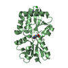
Putative L-Cystine ABC transporter from Listeria monocytogenes
Components
Lmo2349 protein
Keywords
TRANSPORT PROTEIN / structural genomics / IDP05245 / L-Cystine / ABC transporter / periplasmic binding protein / NIAID / National Institute of Allergy and Infectious Diseases / Center for Structural Genomics of Infectious Diseases / CSGID
Function / homology
Function and homology information
Bacterial extracellular solute-binding proteins, family 3 / Solute-binding protein family 3/N-terminal domain of MltF / Bacterial periplasmic substrate-binding proteins / Periplasmic binding protein-like II / D-Maltodextrin-Binding Protein; domain 2 / Prokaryotic membrane lipoprotein lipid attachment site profile. / 3-Layer(aba) Sandwich / Alpha Beta Similarity search - Domain/homology
In the structure databanks used in Yorodumi, some data are registered as the other names, "COVID-19 virus" and "2019-nCoV". Here are the details of the virus and the list of structure data.
Jan 31, 2019. EMDB accession codes are about to change! (news from PDBe EMDB page)
EMDB accession codes are about to change! (news from PDBe EMDB page)
The allocation of 4 digits for EMDB accession codes will soon come to an end. Whilst these codes will remain in use, new EMDB accession codes will include an additional digit and will expand incrementally as the available range of codes is exhausted. The current 4-digit format prefixed with “EMD-” (i.e. EMD-XXXX) will advance to a 5-digit format (i.e. EMD-XXXXX), and so on. It is currently estimated that the 4-digit codes will be depleted around Spring 2019, at which point the 5-digit format will come into force.
The EM Navigator/Yorodumi systems omit the EMD- prefix.
Related info.:Q: What is EMD? / ID/Accession-code notation in Yorodumi/EM Navigator
Yorodumi is a browser for structure data from EMDB, PDB, SASBDB, etc.
This page is also the successor to EM Navigator detail page, and also detail information page/front-end page for Omokage search.
The word "yorodu" (or yorozu) is an old Japanese word meaning "ten thousand". "mi" (miru) is to see.
Related info.:EMDB / PDB / SASBDB / Comparison of 3 databanks / Yorodumi Search / Aug 31, 2016. New EM Navigator & Yorodumi / Yorodumi Papers / Jmol/JSmol / Function and homology information / Changes in new EM Navigator and Yorodumi
 Movie
Movie Controller


 Open data
 Basic information
 Components
 Keywords
 Function and homology information
 Listeria monocytogenes (bacteria)
 X-RAY DIFFRACTION /
  Authors
 Citation
 Structure visualization
 Downloads & links
 Other downloads

 PDBj
PDBj


 Assembly
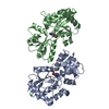


 Mass: 35.453 Da / Num. of mol.: 2 / Source method: obtained synthetically / Formula: Cl
 Mass: 22.990 Da / Num. of mol.: 2 / Source method: obtained synthetically / Formula: Na
 Type: L-peptide linking / Mass: 156.162 Da / Num. of mol.: 2 / Source method: obtained synthetically / Formula: C6H10N3O2
 Type: L-peptide linking / Mass: 137.158 Da / Num. of mol.: 2 / Source method: obtained synthetically / Formula: C3H7NO3S
 Sample preparation
 / Beamline: 19-ID / Wavelength: 0.9792 Å
 Processing