 Movie
Movie Controller

[English] 日本語
 Yorodumi
- PDB-4foo: Crystal Structure of Shikimate Dehydrogenase (aroE) Q237K Mutant ... -
+ Open data

- Basic information
| Entry | Database: PDB / ID: 4foo | ||||||
|---|---|---|---|---|---|---|---|
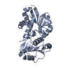
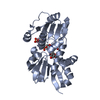
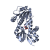
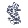
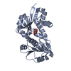
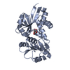
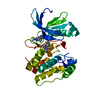
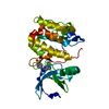
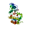
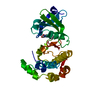
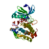
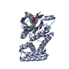
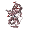
| Title | Crystal Structure of Shikimate Dehydrogenase (aroE) Q237K Mutant from Helicobacter pylori | ||||||
 Components Components | Shikimate dehydrogenase | ||||||
 Keywords Keywords | OXIDOREDUCTASE / shikimate / dehydrogenase / NADP Binding | ||||||
| Function / homology |  Function and homology information Function and homology informationshikimate dehydrogenase (NADP+) / shikimate 3-dehydrogenase (NADP+) activity / shikimate metabolic process / chorismate biosynthetic process / aromatic amino acid biosynthetic process / amino acid biosynthetic process / NADP binding / cytosol Similarity search - Function | ||||||
| Biological species |   Helicobacter pylori (bacteria) Helicobacter pylori (bacteria) | ||||||
| Method |  X-RAY DIFFRACTION / SYNCHROTRON / MOLECULAR REPLACEMENT / Resolution: 2.55 Å X-RAY DIFFRACTION / SYNCHROTRON / MOLECULAR REPLACEMENT / Resolution: 2.55 Å | ||||||
 Authors Authors | Cheng, W.C. / Chen, T.J. / Wang, W.C. | ||||||
| History |
|
- Structure visualization
| Structure viewer | Molecule: MolmilJmol/JSmol |
|---|
- Downloads & links
-Download
| PDBx/mmCIF format | 4foo.cif.gz | 111.2 KB | Display | PDBx/mmCIF format |
|---|---|---|---|---|
| PDB format | pdb4foo.ent.gz | 86.4 KB | Display | PDB format |
| PDBx/mmJSON format | 4foo.json.gz | Tree view | PDBx/mmJSON format | |
| Others |  Other downloads Other downloads |
-Validation report
| Arichive directory | https://data.pdbj.org/pub/pdb/validation_reports/fo/4fooftp://data.pdbj.org/pub/pdb/validation_reports/fo/4foo | HTTPS FTP |
|---|
-Related structure data
| Related structure data | 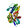 3phhS S: Starting model for refinement |
|---|---|
| Similar structure data |


-Links
 PDBj
PDBj- Assembly
| Deposited unit | 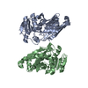
| ||||||||
|---|---|---|---|---|---|---|---|---|---|
| 1 | 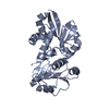
| ||||||||
| 2 | 
| ||||||||
| Unit cell |
|
-Components
| #1: Protein | Mass: 30382.283 Da / Num. of mol.: 2 / Mutation: Q237K Source method: isolated from a genetically manipulated source Source: (gene. exp.) Helicobacter pylori (bacteria) / Strain: 26695 / Gene: aroE, HP_1249 / Plasmid: pET28a / Production host: References: UniProt: P56119, shikimate dehydrogenase (NADP+) #2: Water | ChemComp-HOH / |  Mass: 18.015 Da / Num. of mol.: 50 / Source method: isolated from a natural source / Formula: H2O Mass: 18.015 Da / Num. of mol.: 50 / Source method: isolated from a natural source / Formula: H2O |
|---|
-Experimental details
-Experiment
| Experiment | Method: X-RAY DIFFRACTION / Number of used crystals: 1 |
|---|
- Sample preparation
| Crystal | Density Matthews: 2.04 Å3/Da / Density % sol: 39.7 % |
|---|---|
| Crystal grow | Temperature: 293 K / Method: vapor diffusion, hanging drop / pH: 7 Details: 0.2M potassium acetate, 0.1M Tris, 23% PEG 3350 , pH 7.0, VAPOR DIFFUSION, HANGING DROP, temperature 293K |
-Data collection
| Diffraction | Mean temperature: 110 K | |||||||||||||||||||||||||||||||||||||||||||||||||||||||||||||||||||||||||||||
|---|---|---|---|---|---|---|---|---|---|---|---|---|---|---|---|---|---|---|---|---|---|---|---|---|---|---|---|---|---|---|---|---|---|---|---|---|---|---|---|---|---|---|---|---|---|---|---|---|---|---|---|---|---|---|---|---|---|---|---|---|---|---|---|---|---|---|---|---|---|---|---|---|---|---|---|---|---|---|
| Diffraction source | Source: SYNCHROTRON / Site: NSRRC  / Beamline: BL13C1 / Wavelength: 1 Å / Beamline: BL13C1 / Wavelength: 1 Å | |||||||||||||||||||||||||||||||||||||||||||||||||||||||||||||||||||||||||||||
| Detector | Type: ADSC QUANTUM 210 / Detector: CCD / Date: May 6, 2012 / Details: mirrors | |||||||||||||||||||||||||||||||||||||||||||||||||||||||||||||||||||||||||||||
| Radiation | Monochromator: Si(111) Bent Monochromator / Protocol: SINGLE WAVELENGTH / Monochromatic (M) / Laue (L): M / Scattering type: x-ray | |||||||||||||||||||||||||||||||||||||||||||||||||||||||||||||||||||||||||||||
| Radiation wavelength | Wavelength: 1 Å / Relative weight: 1 | |||||||||||||||||||||||||||||||||||||||||||||||||||||||||||||||||||||||||||||
| Reflection | Resolution: 2.55→30 Å / Num. obs: 16286 / % possible obs: 99.8 % / Redundancy: 3.9 % / Rmerge(I) obs: 0.085 / Χ2: 1.048 / Net I/σ(I): 10.1 | |||||||||||||||||||||||||||||||||||||||||||||||||||||||||||||||||||||||||||||
| Reflection shell | Diffraction-ID: 1
|
- Processing
| Software |
| |||||||||||||||||||||||||||||||||||||||||||||||||||||||||||||||||
|---|---|---|---|---|---|---|---|---|---|---|---|---|---|---|---|---|---|---|---|---|---|---|---|---|---|---|---|---|---|---|---|---|---|---|---|---|---|---|---|---|---|---|---|---|---|---|---|---|---|---|---|---|---|---|---|---|---|---|---|---|---|---|---|---|---|---|
| Refinement | Method to determine structure: MOLECULAR REPLACEMENT Starting model: PDB ENTRY 3PHH Resolution: 2.55→30 Å / Cor.coef. Fo:Fc: 0.933 / Cor.coef. Fo:Fc free: 0.907 / Occupancy max: 1 / Occupancy min: 1 / SU B: 9.159 / SU ML: 0.204 / Cross valid method: THROUGHOUT / σ(F): 0 / ESU R Free: 0.311 / Stereochemistry target values: MAXIMUM LIKELIHOOD Details: HYDROGENS HAVE BEEN ADDED IN THE RIDING POSITIONS U VALUES : REFINED INDIVIDUALLY
| |||||||||||||||||||||||||||||||||||||||||||||||||||||||||||||||||
| Solvent computation | Ion probe radii: 0.8 Å / Shrinkage radii: 0.8 Å / VDW probe radii: 1.4 Å / Solvent model: MASK | |||||||||||||||||||||||||||||||||||||||||||||||||||||||||||||||||
| Displacement parameters | Biso max: 62.6 Å2 / Biso mean: 26.2603 Å2 / Biso min: 2 Å2
| |||||||||||||||||||||||||||||||||||||||||||||||||||||||||||||||||
| Refinement step | Cycle: LAST / Resolution: 2.55→30 Å
| |||||||||||||||||||||||||||||||||||||||||||||||||||||||||||||||||
| Refine LS restraints |
| |||||||||||||||||||||||||||||||||||||||||||||||||||||||||||||||||
| LS refinement shell | Resolution: 2.55→2.616 Å / Total num. of bins used: 20
|
