 Movie
Movie Controller


+ Open data

- Basic information
| Entry | Database: PDB / ID: 7cgw | ||||||
|---|---|---|---|---|---|---|---|
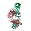
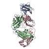
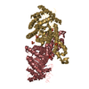
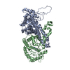
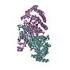
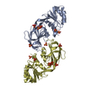
| Title | Complex structure of PD-1 and tislelizumab Fab | ||||||
 Components Components |
| ||||||
 Keywords Keywords | ANTITUMOR PROTEIN / Tislelizumab / PD-1 / antibody | ||||||
| Function / homology |  Function and homology information Function and homology informationregulatory T cell apoptotic process / negative regulation of tolerance induction / negative regulation of immune response / negative regulation of T cell mediated immune response to tumor cell / positive regulation of T cell apoptotic process / B cell apoptotic process / negative regulation of B cell apoptotic process / negative regulation of T cell activation / humoral immune response / Co-inhibition by PD-1 ...regulatory T cell apoptotic process / negative regulation of tolerance induction / negative regulation of immune response / negative regulation of T cell mediated immune response to tumor cell / positive regulation of T cell apoptotic process / B cell apoptotic process / negative regulation of B cell apoptotic process / negative regulation of T cell activation / humoral immune response / Co-inhibition by PD-1 / negative regulation of T cell receptor signaling pathway / regulation of immune response / negative regulation of inflammatory response / transmembrane signaling receptor activity / signaling receptor activity / Potential therapeutics for SARS / adaptive immune response / external side of plasma membrane / apoptotic process / plasma membrane Similarity search - Function | ||||||
| Biological species |  Homo sapiens (human) Homo sapiens (human) | ||||||
| Method |  X-RAY DIFFRACTION / SYNCHROTRON / MOLECULAR REPLACEMENT / Resolution: 3.2 Å X-RAY DIFFRACTION / SYNCHROTRON / MOLECULAR REPLACEMENT / Resolution: 3.2 Å | ||||||
 Authors Authors | Hong, Y. / Feng, Y.C. / Liu, Y. | ||||||
 Citation Citation | Journal: Febs Open Bio / Year: 2021 Title: Tislelizumab uniquely binds to the CC' loop of PD-1 with slow-dissociated rate and complete PD-L1 blockage. Authors: Hong, Y. / Feng, Y. / Sun, H. / Zhang, B. / Wu, H. / Zhu, Q. / Li, Y. / Zhang, T. / Zhang, Y. / Cui, X. / Li, Z. / Song, X. / Li, K. / Liu, M. / Liu, Y. | ||||||
| History |
|
- Structure visualization
| Structure viewer | Molecule: MolmilJmol/JSmol |
|---|
- Downloads & links
-Download
| PDBx/mmCIF format | 7cgw.cif.gz | 242.4 KB | Display | PDBx/mmCIF format |
|---|---|---|---|---|
| PDB format | pdb7cgw.ent.gz | 178.4 KB | Display | PDB format |
| PDBx/mmJSON format | 7cgw.json.gz | Tree view | PDBx/mmJSON format | |
| Others |  Other downloads Other downloads |
-Validation report
| Arichive directory | https://data.pdbj.org/pub/pdb/validation_reports/cg/7cgwftp://data.pdbj.org/pub/pdb/validation_reports/cg/7cgw | HTTPS FTP |
|---|
-Related structure data
| Similar structure data |
|---|


-Links
 PDBj
PDBj


- Assembly
| Deposited unit | 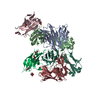
| ||||||||||||
|---|---|---|---|---|---|---|---|---|---|---|---|---|---|
| 1 | 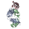
| ||||||||||||
| 2 | 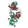
| ||||||||||||
| Unit cell |
|
-Components
| #1: Antibody | Mass: 24477.336 Da / Num. of mol.: 2 Source method: isolated from a genetically manipulated source Source: (gene. exp.) Homo sapiens (human) / Production host: Homo sapiens (human)#2: Antibody | Mass: 23549.080 Da / Num. of mol.: 2 Source method: isolated from a genetically manipulated source Source: (gene. exp.) Homo sapiens (human) / Production host: Homo sapiens (human)#3: Protein | Mass: 16979.893 Da / Num. of mol.: 2 Source method: isolated from a genetically manipulated source Source: (gene. exp.) Homo sapiens (human) / Gene: PDCD1, PD1 / Production host: Homo sapiens (human) / References: UniProt: Q15116#4: Sugar | ChemComp-NAG /   Type: D-saccharide, beta linking / Mass: 221.208 Da / Num. of mol.: 7 Type: D-saccharide, beta linking / Mass: 221.208 Da / Num. of mol.: 7Source method: isolated from a genetically manipulated source Formula: C8H15NO6 / Feature type: SUBJECT OF INVESTIGATION Has ligand of interest | Y | Has protein modification | Y | |
|---|
-Experimental details
-Experiment
| Experiment | Method: X-RAY DIFFRACTION / Number of used crystals: 1 |
|---|
- Sample preparation
| Crystal | Density Matthews: 3.5 Å3/Da / Density % sol: 64.9 % |
|---|---|
| Crystal grow | Temperature: 293 K / Method: vapor diffusion, sitting drop / pH: 4 / Details: 0.1M Citric acid, pH4.0, 1M LiCl and 20% PEG6000 |
-Data collection
| Diffraction | Mean temperature: 93 K / Serial crystal experiment: N |
|---|---|
| Diffraction source | Source: SYNCHROTRON / Site: SSRF  / Beamline: BL17U1 / Wavelength: 0.979 Å / Beamline: BL17U1 / Wavelength: 0.979 Å |
| Detector | Type: DECTRIS EIGER X 16M / Detector: PIXEL / Date: Jun 20, 2018 |
| Radiation | Protocol: SINGLE WAVELENGTH / Monochromatic (M) / Laue (L): M / Scattering type: x-ray |
| Radiation wavelength | Wavelength: 0.979 Å / Relative weight: 1 |
| Reflection | Resolution: 3.2→101.88 Å / Num. obs: 30470 / % possible obs: 99.9 % / Redundancy: 11.4 % / Biso Wilson estimate: 95.76 Å2 / CC1/2: 0.994 / Net I/σ(I): 11.8 |
| Reflection shell | Resolution: 3.2→3.37 Å / Mean I/σ(I) obs: 2.4 / Num. unique obs: 4399 / CC1/2: 0.673 |
- Processing
| Software |
| |||||||||||||||||||||||||||||||||||||||||||||||||||||||||||||||||||||||||||||||||||||||||||||||||||||||||
|---|---|---|---|---|---|---|---|---|---|---|---|---|---|---|---|---|---|---|---|---|---|---|---|---|---|---|---|---|---|---|---|---|---|---|---|---|---|---|---|---|---|---|---|---|---|---|---|---|---|---|---|---|---|---|---|---|---|---|---|---|---|---|---|---|---|---|---|---|---|---|---|---|---|---|---|---|---|---|---|---|---|---|---|---|---|---|---|---|---|---|---|---|---|---|---|---|---|---|---|---|---|---|---|---|---|---|
| Refinement | Method to determine structure: MOLECULAR REPLACEMENT Starting model: Tislelizumab Fab Resolution: 3.2→101.88 Å / SU ML: 0.4264 / Cross valid method: FREE R-VALUE / σ(F): 1.33 / Phase error: 27.2957 Stereochemistry target values: GeoStd + Monomer Library + CDL v1.2
| |||||||||||||||||||||||||||||||||||||||||||||||||||||||||||||||||||||||||||||||||||||||||||||||||||||||||
| Solvent computation | Shrinkage radii: 0.9 Å / VDW probe radii: 1.11 Å / Solvent model: FLAT BULK SOLVENT MODEL | |||||||||||||||||||||||||||||||||||||||||||||||||||||||||||||||||||||||||||||||||||||||||||||||||||||||||
| Displacement parameters | Biso mean: 104.54 Å2 | |||||||||||||||||||||||||||||||||||||||||||||||||||||||||||||||||||||||||||||||||||||||||||||||||||||||||
| Refinement step | Cycle: LAST / Resolution: 3.2→101.88 Å
| |||||||||||||||||||||||||||||||||||||||||||||||||||||||||||||||||||||||||||||||||||||||||||||||||||||||||
| Refine LS restraints |
| |||||||||||||||||||||||||||||||||||||||||||||||||||||||||||||||||||||||||||||||||||||||||||||||||||||||||
| LS refinement shell |
|
