 Movie
Movie Controller

[English] 日本語
 Yorodumi
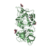
- PDB-4akr: Crystal Structure of the cytoplasmic actin capping protein Cap32_... -
+ Open data

- Basic information
| Entry | Database: PDB / ID: 4akr | ||||||
|---|---|---|---|---|---|---|---|
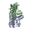
| Title | Crystal Structure of the cytoplasmic actin capping protein Cap32_34 from Dictyostelium discoideum | ||||||
 Components Components |
| ||||||
 Keywords Keywords | ACTIN-BINDING PROTEIN | ||||||
| Function / homology |  Function and homology information Function and homology informationFactors involved in megakaryocyte development and platelet production / COPI-mediated anterograde transport / F-actin capping protein complex / barbed-end actin filament capping / cortical actin cytoskeleton / cortical cytoskeleton / response to bacterium / cell morphogenesis / actin filament binding / actin cytoskeleton organization ...Factors involved in megakaryocyte development and platelet production / COPI-mediated anterograde transport / F-actin capping protein complex / barbed-end actin filament capping / cortical actin cytoskeleton / cortical cytoskeleton / response to bacterium / cell morphogenesis / actin filament binding / actin cytoskeleton organization / lipid binding / cytosol Similarity search - Function | ||||||
| Biological species |  | ||||||
| Method |  X-RAY DIFFRACTION / SYNCHROTRON / MOLECULAR REPLACEMENT / Resolution: 2.2 Å X-RAY DIFFRACTION / SYNCHROTRON / MOLECULAR REPLACEMENT / Resolution: 2.2 Å | ||||||
 Authors Authors | Eckert, C. / Goretzki, A. / Faberova, M. / Kollmar, M. | ||||||
 Citation Citation | Journal: Bmc Struct.Biol. / Year: 2012 Title: Conservation and Divergence between Cytoplasmic and Muscle-Specific Actin Capping Proteins: Insights from the Crystal Structure of Cytoplasmic CAP32/34 from Dictyostelium Discoideum. Authors: Eckert, C. / Goretzki, A. / Faberova, M. / Kollmar, M. | ||||||
| History |
|
- Structure visualization
| Structure viewer | Molecule: MolmilJmol/JSmol |
|---|
- Downloads & links
-Download
| PDBx/mmCIF format | 4akr.cif.gz | 213.9 KB | Display | PDBx/mmCIF format |
|---|---|---|---|---|
| PDB format | pdb4akr.ent.gz | 171.5 KB | Display | PDB format |
| PDBx/mmJSON format | 4akr.json.gz | Tree view | PDBx/mmJSON format | |
| Others |  Other downloads Other downloads |
-Validation report
| Arichive directory | https://data.pdbj.org/pub/pdb/validation_reports/ak/4akrftp://data.pdbj.org/pub/pdb/validation_reports/ak/4akr | HTTPS FTP |
|---|
-Related structure data
| Related structure data | 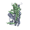 1iznS S: Starting model for refinement |
|---|---|
| Similar structure data |


-Links
 PDBj
PDBj
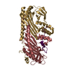
- Assembly
| Deposited unit | 
| ||||||||
|---|---|---|---|---|---|---|---|---|---|
| 1 | 
| ||||||||
| 2 | 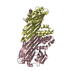
| ||||||||
| Unit cell |
|
-Components
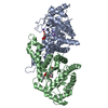
| #1: Protein | Mass: 31194.803 Da / Num. of mol.: 2 Source method: isolated from a genetically manipulated source Source: (gene. exp.) Description: CDNA FROM THE DICTYOSTELIUM CDNA PROJECT IN JAPAN AND THE JAPANESE NATIONAL BIORESOURCE PROJECT Production host:  #2: Protein | Mass: 32873.859 Da / Num. of mol.: 2 / Fragment: RESIDUES 2-272 Source method: isolated from a genetically manipulated source Source: (gene. exp.) Description: CDNA FROM THE DICTYOSTELIUM CDNA PROJECT IN JAPAN AND THE JAPANESE NATIONAL BIORESOURCE PROJECT Production host: #3: Water | ChemComp-HOH / |  Mass: 18.015 Da / Num. of mol.: 330 / Source method: isolated from a natural source / Formula: H2O Mass: 18.015 Da / Num. of mol.: 330 / Source method: isolated from a natural source / Formula: H2O |
|---|
-Experimental details
-Experiment
| Experiment | Method: X-RAY DIFFRACTION / Number of used crystals: 1 |
|---|
- Sample preparation
| Crystal | Density Matthews: 2.27 Å3/Da / Density % sol: 46 % / Description: NONE |
|---|---|
| Crystal grow | pH: 7.5 / Details: 100 MM HEPES PH 7.5, 17% (W/V) PEG 8000 |
-Data collection
| Diffraction | Mean temperature: 100 K |
|---|---|
| Diffraction source | Source: SYNCHROTRON / Site: ESRF  / Beamline: ID23-2 / Wavelength: 0.8726 / Beamline: ID23-2 / Wavelength: 0.8726 |
| Detector | Type: MARMOSAIC 225 mm CCD / Detector: CCD / Date: May 4, 2010 |
| Radiation | Protocol: SINGLE WAVELENGTH / Monochromatic (M) / Laue (L): M / Scattering type: x-ray |
| Radiation wavelength | Wavelength: 0.8726 Å / Relative weight: 1 |
| Reflection | Resolution: 2.2→50 Å / Num. obs: 60185 / % possible obs: 99.8 % / Observed criterion σ(I): 3 / Redundancy: 6.1 % / Rmerge(I) obs: 0.15 / Net I/σ(I): 14.15 |
| Reflection shell | Resolution: 2.2→2.3 Å / Redundancy: 6.1 % / Rmerge(I) obs: 0.84 / Mean I/σ(I) obs: 3.83 / % possible all: 99.6 |
- Processing
| Software |
| ||||||||||||||||||||||||||||||||||||||||||||||||||||||||||||
|---|---|---|---|---|---|---|---|---|---|---|---|---|---|---|---|---|---|---|---|---|---|---|---|---|---|---|---|---|---|---|---|---|---|---|---|---|---|---|---|---|---|---|---|---|---|---|---|---|---|---|---|---|---|---|---|---|---|---|---|---|---|
| Refinement | Method to determine structure: MOLECULAR REPLACEMENT Starting model: PDB ENTRY 1IZN Resolution: 2.2→50 Å / Cross valid method: THROUGHOUT / σ(F): 3 / Stereochemistry target values: MAXIMUM LIKELIHOOD
| ||||||||||||||||||||||||||||||||||||||||||||||||||||||||||||
| Displacement parameters | Biso mean: 15.4 Å2 | ||||||||||||||||||||||||||||||||||||||||||||||||||||||||||||
| Refinement step | Cycle: LAST / Resolution: 2.2→50 Å
| ||||||||||||||||||||||||||||||||||||||||||||||||||||||||||||
| Refine LS restraints |
|
