 Movie
Movie Controller

[English] 日本語
 Yorodumi
- PDB-3lk2: Crystal structure of CapZ bound to the uncapping motif from CARMIL -
+ Open data

- Basic information
| Entry | Database: PDB / ID: 3lk2 | ||||||
|---|---|---|---|---|---|---|---|
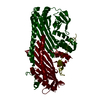
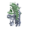
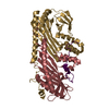
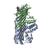
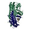
| Title | Crystal structure of CapZ bound to the uncapping motif from CARMIL | ||||||
 Components Components |
| ||||||
 Keywords Keywords | PROTEIN BINDING / CapZ / CARMIL / actin filaments / uncapping / actin-filament regulators / protein-protein complex / Actin capping / Actin-binding / Cytoskeleton | ||||||
| Function / homology |  Function and homology information Function and homology informationbarbed-end actin filament uncapping / positive regulation of lamellipodium organization / negative regulation of barbed-end actin filament capping / Advanced glycosylation endproduct receptor signaling / macropinosome / negative regulation of filopodium assembly / actin filament network formation / F-actin capping protein complex / WASH complex / macropinocytosis ...barbed-end actin filament uncapping / positive regulation of lamellipodium organization / negative regulation of barbed-end actin filament capping / Advanced glycosylation endproduct receptor signaling / macropinosome / negative regulation of filopodium assembly / actin filament network formation / F-actin capping protein complex / WASH complex / macropinocytosis / regulation of Arp2/3 complex-mediated actin nucleation / urate metabolic process / cell junction assembly / ruffle organization / barbed-end actin filament capping / regulation of lamellipodium assembly / sperm head-tail coupling apparatus / lamellipodium assembly / positive regulation of actin filament polymerization / filamentous actin / cell leading edge / cortical cytoskeleton / brush border / positive regulation of stress fiber assembly / positive regulation of substrate adhesion-dependent cell spreading / actin filament organization / cell morphogenesis / Schaffer collateral - CA1 synapse / Z disc / actin filament binding / cell migration / lamellipodium / Factors involved in megakaryocyte development and platelet production / actin cytoskeleton organization / postsynaptic density / positive regulation of cell migration / protein-containing complex binding / extracellular exosome / membrane / plasma membrane / cytosol Similarity search - Function | ||||||
| Biological species |  | ||||||
| Method |  X-RAY DIFFRACTION / SYNCHROTRON / MOLECULAR REPLACEMENT / Resolution: 2.2 Å X-RAY DIFFRACTION / SYNCHROTRON / MOLECULAR REPLACEMENT / Resolution: 2.2 Å | ||||||
 Authors Authors | Hernandez-Valladares, M. / Kim, T. / Kannan, B. / Tung, A. / Aguda, A.H. / Larsson, M. / Cooper, J.A. / Robinson, R.C. | ||||||
 Citation Citation | Journal: Nat.Struct.Mol.Biol. / Year: 2010 Title: Structural characterization of a capping protein interaction motif defines a family of actin filament regulators. Authors: Hernandez-Valladares, M. / Kim, T. / Kannan, B. / Tung, A. / Aguda, A.H. / Larsson, M. / Cooper, J.A. / Robinson, R.C. | ||||||
| History |
|
- Structure visualization
| Structure viewer | Molecule: MolmilJmol/JSmol |
|---|
- Downloads & links
-Download
| PDBx/mmCIF format | 3lk2.cif.gz | 127 KB | Display | PDBx/mmCIF format |
|---|---|---|---|---|
| PDB format | pdb3lk2.ent.gz | 97.5 KB | Display | PDB format |
| PDBx/mmJSON format | 3lk2.json.gz | Tree view | PDBx/mmJSON format | |
| Others |  Other downloads Other downloads |
-Validation report
| Arichive directory | https://data.pdbj.org/pub/pdb/validation_reports/lk/3lk2ftp://data.pdbj.org/pub/pdb/validation_reports/lk/3lk2 | HTTPS FTP |
|---|
-Related structure data
| Related structure data |  3lk3C 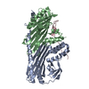 3lk4C 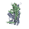 1iznS S: Starting model for refinement C: citing same article ( |
|---|---|
| Similar structure data |


-Links
 PDBj
PDBj


- Assembly
| Deposited unit | 
| ||||||||
|---|---|---|---|---|---|---|---|---|---|
| 1 |
| ||||||||
| Unit cell |
|
-Components
| #1: Protein | Mass: 33001.789 Da / Num. of mol.: 1 Source method: isolated from a genetically manipulated source Source: (gene. exp.)  |
|---|---|
| #2: Protein | Mass: 27315.877 Da / Num. of mol.: 1 Source method: isolated from a genetically manipulated source Source: (gene. exp.) |
| #3: Protein | Mass: 5920.755 Da / Num. of mol.: 1 / Fragment: CPI, capping protein interacting motif / Source method: obtained synthetically Details: The peptide was chemically synthesized. The sequence of the peptide is naturally found in Homo sapiens (human) References: UniProt: Q5VZK9 |
| #4: Water | ChemComp-HOH /  Mass: 18.015 Da / Num. of mol.: 260 / Source method: isolated from a natural source / Formula: H2O Mass: 18.015 Da / Num. of mol.: 260 / Source method: isolated from a natural source / Formula: H2O |
-Experimental details
-Experiment
| Experiment | Method: X-RAY DIFFRACTION / Number of used crystals: 1 |
|---|
- Sample preparation
| Crystal | Density Matthews: 2.29 Å3/Da / Density % sol: 46.35 % |
|---|---|
| Crystal grow | Temperature: 277 K / Method: vapor diffusion, sitting drop / pH: 6.5 Details: 10% w/v PEG 20000, 100mM MES, pH 6.5, VAPOR DIFFUSION, SITTING DROP, temperature 277K |
-Data collection
| Diffraction | Mean temperature: 105 K |
|---|---|
| Diffraction source | Source: SYNCHROTRON / Site: NSRRC  / Beamline: BL13B1 / Wavelength: 1 Å / Beamline: BL13B1 / Wavelength: 1 Å |
| Detector | Type: ADSC QUANTUM 315 / Detector: CCD / Date: Nov 14, 2008 / Details: phosphor screen, fiber-optic taper and CCD chip |
| Radiation | Monochromator: double-crystal / Protocol: SINGLE WAVELENGTH / Monochromatic (M) / Laue (L): M / Scattering type: x-ray |
| Radiation wavelength | Wavelength: 1 Å / Relative weight: 1 |
| Reflection | Resolution: 2.2→20 Å / Num. all: 31702 / Num. obs: 31671 / % possible obs: 99.9 % / Observed criterion σ(F): 0 / Observed criterion σ(I): 0 / Redundancy: 6.9 % / Biso Wilson estimate: 40.9 Å2 / Rmerge(I) obs: 0.063 / Rsym value: 0.063 / Net I/σ(I): 20.4 |
| Reflection shell | Resolution: 2.2→2.3 Å / Redundancy: 6.9 % / Rmerge(I) obs: 0.37 / Mean I/σ(I) obs: 8.1 / Num. unique all: 1514 / Rsym value: 0.37 / % possible all: 100 |
- Processing
| Software |
| ||||||||||||||||||||||||||||||||||||||||||||||||||||||||||||||||||||||||||||||||||||||||||||||||||||
|---|---|---|---|---|---|---|---|---|---|---|---|---|---|---|---|---|---|---|---|---|---|---|---|---|---|---|---|---|---|---|---|---|---|---|---|---|---|---|---|---|---|---|---|---|---|---|---|---|---|---|---|---|---|---|---|---|---|---|---|---|---|---|---|---|---|---|---|---|---|---|---|---|---|---|---|---|---|---|---|---|---|---|---|---|---|---|---|---|---|---|---|---|---|---|---|---|---|---|---|---|---|
| Refinement | Method to determine structure: MOLECULAR REPLACEMENT Starting model: PDB ENTRY 1IZN Resolution: 2.2→19.71 Å / Cor.coef. Fo:Fc: 0.948 / Cor.coef. Fo:Fc free: 0.912 / SU B: 14.784 / SU ML: 0.17 / TLS residual ADP flag: LIKELY RESIDUAL / Isotropic thermal model: Isotropic / Cross valid method: THROUGHOUT / ESU R: 0.298 / ESU R Free: 0.235 / Stereochemistry target values: MAXIMUM LIKELIHOOD
| ||||||||||||||||||||||||||||||||||||||||||||||||||||||||||||||||||||||||||||||||||||||||||||||||||||
| Solvent computation | Ion probe radii: 0.8 Å / Shrinkage radii: 0.8 Å / VDW probe radii: 1.4 Å / Solvent model: MASK | ||||||||||||||||||||||||||||||||||||||||||||||||||||||||||||||||||||||||||||||||||||||||||||||||||||
| Displacement parameters | Biso mean: 21.8 Å2 | ||||||||||||||||||||||||||||||||||||||||||||||||||||||||||||||||||||||||||||||||||||||||||||||||||||
| Refinement step | Cycle: LAST / Resolution: 2.2→19.71 Å
| ||||||||||||||||||||||||||||||||||||||||||||||||||||||||||||||||||||||||||||||||||||||||||||||||||||
| Refine LS restraints |
| ||||||||||||||||||||||||||||||||||||||||||||||||||||||||||||||||||||||||||||||||||||||||||||||||||||
| LS refinement shell | Resolution: 2.2→2.256 Å / Total num. of bins used: 20
| ||||||||||||||||||||||||||||||||||||||||||||||||||||||||||||||||||||||||||||||||||||||||||||||||||||
| Refinement TLS params. | Method: refined / Refine-ID: X-RAY DIFFRACTION
| ||||||||||||||||||||||||||||||||||||||||||||||||||||||||||||||||||||||||||||||||||||||||||||||||||||
| Refinement TLS group |
|
