 Movie
Movie Controller

[English] 日本語
 Yorodumi
- PDB-2zfc: X-ray crystal structure of an engineered N-terminal HIV-1 GP41 tr... -
+ Open data

- Basic information
| Entry | Database: PDB / ID: 2zfc | ||||||
|---|---|---|---|---|---|---|---|
| Title | X-ray crystal structure of an engineered N-terminal HIV-1 GP41 trimer with enhanced stability and potency | ||||||
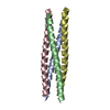
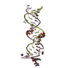
 Components Components | HIV-1 GP41 | ||||||
 Keywords Keywords | VIRAL PROTEIN / hiv-1 / gp41 | ||||||
| Function / homology | Single alpha-helices involved in coiled-coils or other helix-helix interfaces - #170 / Single alpha-helices involved in coiled-coils or other helix-helix interfaces / Up-down Bundle / Mainly Alpha Function and homology information Function and homology information | ||||||
| Method |  X-RAY DIFFRACTION / SAD / Resolution: 1.5 Å X-RAY DIFFRACTION / SAD / Resolution: 1.5 Å | ||||||
 Authors Authors | Dwyer, J.J. / Wilson, K.L. / Martin, K. / Seedorff, J.E. / Hasan, A. / Kim, H. | ||||||
 Citation Citation | Journal: Protein Sci. / Year: 2008 Title: Design of an engineered N-terminal HIV-1 gp41 trimer with enhanced stability and potency Authors: Dwyer, J.J. / Wilson, K.L. / Martin, K. / Seedorff, J.E. / Hasan, A. / Medinas, R.J. / Davison, D.K. / Feese, M.D. / Richter, H.T. / Kim, H. / Matthews, T.J. / Delmedico, M.K. | ||||||
| History |
|
- Structure visualization
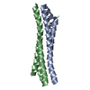
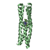
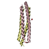
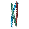
| Structure viewer | Molecule: MolmilJmol/JSmol |
|---|
- Downloads & links
-Download
| PDBx/mmCIF format | 2zfc.cif.gz | 39.5 KB | Display | PDBx/mmCIF format |
|---|---|---|---|---|
| PDB format | pdb2zfc.ent.gz | 28.7 KB | Display | PDB format |
| PDBx/mmJSON format | 2zfc.json.gz | Tree view | PDBx/mmJSON format | |
| Others |  Other downloads Other downloads |
-Validation report
| Arichive directory | https://data.pdbj.org/pub/pdb/validation_reports/zf/2zfcftp://data.pdbj.org/pub/pdb/validation_reports/zf/2zfc | HTTPS FTP |
|---|
-Related structure data
| Similar structure data |
|---|


-Links
 PDBj
PDBj
- Assembly
| Deposited unit | 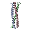
| ||||||||
|---|---|---|---|---|---|---|---|---|---|
| 1 |
| ||||||||
| Unit cell |
|
-Components
| #1: Protein/peptide | Mass: 5558.464 Da / Num. of mol.: 3 / Source method: obtained synthetically / Details: synthetic peptide #2: Water | ChemComp-HOH / |  Mass: 18.015 Da / Num. of mol.: 185 / Source method: isolated from a natural source / Formula: H2O Mass: 18.015 Da / Num. of mol.: 185 / Source method: isolated from a natural source / Formula: H2O |
|---|
-Experimental details
-Experiment
| Experiment | Method: X-RAY DIFFRACTION / Number of used crystals: 1 |
|---|
- Sample preparation
| Crystal | Density Matthews: 2.78 Å3/Da / Density % sol: 55.74 % |
|---|---|
| Crystal grow | Temperature: 289 K / Method: vapor diffusion, sitting drop / pH: 5.6 Details: 10%(v/v) iso-propanol, 0.1M Na Citrate pH 5.6, 10%(w/v) PEG 4000, VAPOR DIFFUSION, SITTING DROP, temperature 289K |
-Data collection
| Diffraction | Mean temperature: 100 K |
|---|---|
| Diffraction source | Source: ROTATING ANODE / Type: RIGAKU RU300 / Wavelength: 1.54 Å |
| Detector | Type: RIGAKU RAXIS IV++ / Detector: IMAGE PLATE / Details: Osmic Blue |
| Radiation | Protocol: SINGLE WAVELENGTH / Monochromatic (M) / Laue (L): M / Scattering type: x-ray |
| Radiation wavelength | Wavelength: 1.54 Å / Relative weight: 1 |
| Reflection | Resolution: 1.5→50 Å / Num. obs: 28563 / % possible obs: 95.5 % / Redundancy: 5 % / Biso Wilson estimate: 24.9 Å2 / Rsym value: 0.046 / Net I/σ(I): 44 |
| Reflection shell | Resolution: 1.5→1.55 Å / Mean I/σ(I) obs: 1.6 / Rsym value: 0.312 / % possible all: 68.6 |
-Phasing
| Phasing | Method: SAD |
|---|
- Processing
| Software |
| ||||||||||||||||||||||||||||
|---|---|---|---|---|---|---|---|---|---|---|---|---|---|---|---|---|---|---|---|---|---|---|---|---|---|---|---|---|---|
| Refinement | Method to determine structure: SAD / Resolution: 1.5→20 Å /
| ||||||||||||||||||||||||||||
| Displacement parameters | Biso mean: 27.739 Å2 | ||||||||||||||||||||||||||||
| Refinement step | Cycle: LAST / Resolution: 1.5→20 Å
| ||||||||||||||||||||||||||||
| Refine LS restraints |
| ||||||||||||||||||||||||||||
| LS refinement shell | Highest resolution: 1.5 Å / Rfactor Rfree: 0.266 / Rfactor Rwork: 0.221 |
