Mass: 18.015 Da / Num. of mol.: 87 / Source method: isolated from a natural source / Formula: H2O
Sequence details
THE N-TERMINAL GLYCINE RESIDUE IS NOT PART OF THE NATIVE SEQUENCE AND DERIVES FROM THE TEV CLEAVAGE ...THE N-TERMINAL GLYCINE RESIDUE IS NOT PART OF THE NATIVE SEQUENCE AND DERIVES FROM THE TEV CLEAVAGE SITE. SEQUENCE USED HERE IS THE ISOFORM-2 OF O95834
-
Experimental details
-
Experiment
Experiment
Method: X-RAY DIFFRACTION / Number of used crystals: 1
-
Sample preparation
Crystal
Density Matthews: 1.86 Å3/Da / Density % sol: 33.7 % / Description: NONE
Crystal grow
Method: vapor diffusion, hanging drop / pH: 6.5 Details: CRYSTALS WERE OBTAINED BY THE HANGING DROP VAPOUR DIFFUSION METHOD USING 100 MM BIS-TRIS-PROPANE PH 6.5, 20% PEG 3350, 200 MM POTASSIUM THIOCYANATE IN THE RESERVOIR.
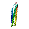
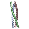
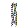
Resolution: 2.154→50.846 Å / SU ML: 0.2 / σ(F): 1.02 / Phase error: 23.5 / Stereochemistry target values: ML Details: IN CHAIN A RESIDUES 14-57 ARE ORDERED. IN CHAIN B RESIDUES 14-58 ARE ORDERED. IN CHAIN C RESIDUES 13-53 ARE ORDERED. IN CHAIN D RESIDUES 13-57 ARE ORDERED. IN CHAIN E RESIDUES 13-52 ARE ...Details: IN CHAIN A RESIDUES 14-57 ARE ORDERED. IN CHAIN B RESIDUES 14-58 ARE ORDERED. IN CHAIN C RESIDUES 13-53 ARE ORDERED. IN CHAIN D RESIDUES 13-57 ARE ORDERED. IN CHAIN E RESIDUES 13-52 ARE ORDERED. IN CHAIN F RESIDUES 13-57 ARE ORDERED.
Rfactor
Num. reflection
% reflection
Rfree
0.2515
696
5 %
Rwork
0.2055
-
-
obs
0.2078
14027
99.67 %
Solvent computation
Shrinkage radii: 0.98 Å / VDW probe radii: 1.2 Å / Solvent model: FLAT BULK SOLVENT MODEL / Bsol: 43.888 Å2 / ksol: 0.378 e/Å3
Displacement parameters
Baniso -1
Baniso -2
Baniso -3
1-
-6.143 Å2
0 Å2
0 Å2
2-
-
10.0468 Å2
0 Å2
3-
-
-
-3.9039 Å2
Refinement step
Cycle: LAST / Resolution: 2.154→50.846 Å
Protein
Nucleic acid
Ligand
Solvent
Total
Num. atoms
1968
0
8
87
2063
Refine LS restraints
Refine-ID
Type
Dev ideal
Number
X-RAY DIFFRACTION
f_bond_d
0.006
1991
X-RAY DIFFRACTION
f_angle_d
0.93
2680
X-RAY DIFFRACTION
f_dihedral_angle_d
17.187
777
X-RAY DIFFRACTION
f_chiral_restr
0.045
329
X-RAY DIFFRACTION
f_plane_restr
0.003
362
LS refinement shell
Resolution (Å)
Rfactor Rfree
Num. reflection Rfree
Rfactor Rwork
Num. reflection Rwork
Refine-ID
% reflection obs (%)
2.1542-2.3205
0.3018
144
0.2138
2593
X-RAY DIFFRACTION
100
2.3205-2.554
0.2559
144
0.2046
2617
X-RAY DIFFRACTION
100
2.554-2.9235
0.2751
130
0.2187
2639
X-RAY DIFFRACTION
99
2.9235-3.6832
0.2486
147
0.1918
2676
X-RAY DIFFRACTION
100
3.6832-50.8596
0.2315
131
0.2079
2806
X-RAY DIFFRACTION
100
Refinement TLS params.
Method: refined / Refine-ID: X-RAY DIFFRACTION
ID
L11 (°2)
L12 (°2)
L13 (°2)
L22 (°2)
L23 (°2)
L33 (°2)
S11 (Å °)
S12 (Å °)
S13 (Å °)
S21 (Å °)
S22 (Å °)
S23 (Å °)
S31 (Å °)
S32 (Å °)
S33 (Å °)
T11 (Å2)
T12 (Å2)
T13 (Å2)
T22 (Å2)
T23 (Å2)
T33 (Å2)
Origin x (Å)
Origin y (Å)
Origin z (Å)
1
4.1249
1.0983
3.7691
2.7935
1.6389
9.1057
-0.0898
-0.1218
0.1097
-0.0575
0.0321
0.1005
-0.1685
-0.3533
0.1268
0.1745
0.0118
0.0439
0.1466
0.0434
0.2342
-1.1788
21.6685
48.0306
2
3.9328
-0.2788
1.6424
1.5038
0.1857
2.6759
0.1525
0.1127
-0.0604
-0.0713
0.0298
-0.0489
0.3529
-0.2631
-0.2653
0.1239
0.0101
0.0352
0.1125
0.0126
0.2266
5.3166
17.4358
47.1587
3
6.1042
-0.1503
4.5165
1.4969
-0.9334
6.4932
0.0338
0.3069
0.0767
0.0658
0.1249
0.1543
-0.3848
0.3033
-0.1294
0.2794
-0.0311
0.0386
0.2025
0.0519
0.3179
2.4572
25.5088
41.7592
4
5.1138
3.4841
-2.3207
5.9317
-3.0982
3.539
-0.1014
0.7377
0.2314
-0.2209
0.3129
0.3449
0.0848
-0.3285
-0.1943
0.2402
0.0704
-0.0557
0.3014
-0.0065
0.1978
16.5496
15.5203
37.3313
5
2.3212
3.2814
0.2098
8.637
-0.9064
2.8784
0.0823
0.0967
0.0317
-0.1329
-0.211
-0.2896
-0.1541
0.081
0.1196
0.1631
0.0454
0.0254
0.2514
0.0166
0.2095
23.7293
15.3563
41.5588
6
3.025
2.7956
-0.7238
10.0437
-2.8898
2.7822
-0.0091
0.2953
0.1821
0.1091
-0.0121
-0.0045
-0.1478
0.0197
-0.0992
0.1255
0.0105
-0.0162
0.2625
-0.0154
0.2245
16.4287
17.4424
44.4217
Refinement TLS group
ID
Refine-ID
Refine TLS-ID
Selection details
1
X-RAY DIFFRACTION
1
CHAINAAND (RESSEQ4:57)
2
X-RAY DIFFRACTION
2
CHAINBAND (RESSEQ13:60)
3
X-RAY DIFFRACTION
3
CHAINCAND (RESSEQ13:60)
4
X-RAY DIFFRACTION
4
CHAINDAND (RESSEQ13:57)
5
X-RAY DIFFRACTION
5
CHAINEAND (RESSEQ13:60)
6
X-RAY DIFFRACTION
6
CHAINFAND (RESSEQ13:60)
+
About Yorodumi
-
News
-
Feb 9, 2022. New format data for meta-information of EMDB entries
New format data for meta-information of EMDB entries
Version 3 of the EMDB header file is now the official format.
The previous official version 1.9 will be removed from the archive.
In the structure databanks used in Yorodumi, some data are registered as the other names, "COVID-19 virus" and "2019-nCoV". Here are the details of the virus and the list of structure data.
Jan 31, 2019. EMDB accession codes are about to change! (news from PDBe EMDB page)
EMDB accession codes are about to change! (news from PDBe EMDB page)
The allocation of 4 digits for EMDB accession codes will soon come to an end. Whilst these codes will remain in use, new EMDB accession codes will include an additional digit and will expand incrementally as the available range of codes is exhausted. The current 4-digit format prefixed with “EMD-” (i.e. EMD-XXXX) will advance to a 5-digit format (i.e. EMD-XXXXX), and so on. It is currently estimated that the 4-digit codes will be depleted around Spring 2019, at which point the 5-digit format will come into force.
The EM Navigator/Yorodumi systems omit the EMD- prefix.
Related info.:Q: What is EMD? / ID/Accession-code notation in Yorodumi/EM Navigator
Yorodumi is a browser for structure data from EMDB, PDB, SASBDB, etc.
This page is also the successor to EM Navigator detail page, and also detail information page/front-end page for Omokage search.
The word "yorodu" (or yorozu) is an old Japanese word meaning "ten thousand". "mi" (miru) is to see.
Related info.:EMDB / PDB / SASBDB / Comparison of 3 databanks / Yorodumi Search / Aug 31, 2016. New EM Navigator & Yorodumi / Yorodumi Papers / Jmol/JSmol / Function and homology information / Changes in new EM Navigator and Yorodumi
 Movie
Movie Controller


 Open data
 Basic information
 Components
 Keywords
 Function and homology information
 HOMO SAPIENS (human)
 X-RAY DIFFRACTION /
  Authors
 Citation
 Structure visualization
 Downloads & links
 Other downloads
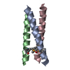

 PDBj
PDBj Assembly
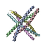


 Mass: 39.098 Da / Num. of mol.: 2 / Source method: obtained synthetically / Formula: K
 Mass: 92.094 Da / Num. of mol.: 1 / Source method: obtained synthetically / Formula: C3H8O3
 Mass: 18.015 Da / Num. of mol.: 87 / Source method: isolated from a natural source / Formula: H2O
 Sample preparation
 / Beamline: I04 / Wavelength: 0.9795
  Processing