[English] 日本語
Yorodumi
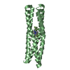
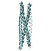
- PDB-3d0t: Structure of the BNB domain of the Hsp70 cochaperone Bag2 -

+
Open data


ID or keywords:

Loading...

-
Basic information

Entry
Database: PDB / ID: 3d0t
TitleStructure of the BNB domain of the Hsp70 cochaperone Bag2
ComponentsBAG family molecular chaperone regulator 2
Keywords
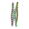
CHAPERONE / 4-helix bundle / Coiled coil
Function / homology
Function and homology information


Regulation of HSF1-mediated heat shock response / adenyl-nucleotide exchange factor activity / positive regulation of protein processing / positive regulation of proteasomal protein catabolic process / protein metabolic process / protein folding chaperone complex / negative regulation of protein ubiquitination / heat shock protein binding / tau protein binding / protein-folding chaperone binding ...Regulation of HSF1-mediated heat shock response / adenyl-nucleotide exchange factor activity / positive regulation of protein processing / positive regulation of proteasomal protein catabolic process / protein metabolic process / protein folding chaperone complex / negative regulation of protein ubiquitination / heat shock protein binding / tau protein binding / protein-folding chaperone binding / transmembrane transporter binding / protein stabilization / axon / ubiquitin protein ligase binding / dendrite / identical protein binding
Similarity search - Function
Methane Monooxygenase Hydroxylase; Chain G, domain 1 - #890 / BAG family molecular chaperone regulator 2 / BAG domain / BAG domain profile. / BAG domains, present in regulator of Hsp70 proteins / Methane Monooxygenase Hydroxylase; Chain G, domain 1 / Up-down Bundle / Mainly Alpha
Similarity search - Domain/homology
BAG family molecular chaperone regulator 2
Similarity search - Component
Biological speciesMus musculus (house mouse)
MethodX-RAY DIFFRACTION / SYNCHROTRON / SAD / Resolution: 2.55 Å
AuthorsXu, Z. / Nix, J.C. / Devlin, K. / Misra, S.
CitationJournal: Nat.Struct.Mol.Biol. / Year: 2008
Title: Structural basis of nucleotide exchange and client binding by the Hsp70 cochaperone Bag2.
Authors: Xu, Z. / Page, R.C. / Gomes, M.M. / Kohli, E. / Nix, J.C. / Herr, A.B. / Patterson, C. / Misra, S.
History
DepositionMay 2, 2008Deposition site: RCSB / Processing site: RCSB
Revision 1.0Nov 25, 2008Provider: repository / Type: Initial release
Revision 1.1Jul 13, 2011Group: Version format compliance
Revision 1.2Feb 21, 2024Group: Data collection / Database references
Category: chem_comp_atom / chem_comp_bond ...chem_comp_atom / chem_comp_bond / database_2 / struct_ref_seq_dif
Item: _database_2.pdbx_DOI / _database_2.pdbx_database_accession / _struct_ref_seq_dif.details

-
Structure visualization

Structure viewerMolecule:
MolmilJmol/JSmol

Downloads & links

-
Assembly

Deposited unit
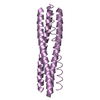
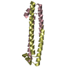
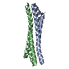
A: BAG family molecular chaperone regulator 2
B: BAG family molecular chaperone regulator 2
C: BAG family molecular chaperone regulator 2
D: BAG family molecular chaperone regulator 2


Theoretical massNumber of molelcules
Total (without water)38,8044
Polymers38,8044
Non-polymers00
Water95553
1
C: BAG family molecular chaperone regulator 2
D: BAG family molecular chaperone regulator 2


Theoretical massNumber of molelcules
Total (without water)19,4022
Polymers19,4022
Non-polymers00
Water362
TypeNameSymmetry operationNumber
identity operation1_555x,y,z1
Buried area3260 Å2
ΔGint-31 kcal/mol
Surface area9220 Å2
MethodPISA
2
A: BAG family molecular chaperone regulator 2
B: BAG family molecular chaperone regulator 2


Theoretical massNumber of molelcules
Total (without water)19,4022
Polymers19,4022
Non-polymers00
Water362
TypeNameSymmetry operationNumber
identity operation1_555x,y,z1
Buried area3190 Å2
ΔGint-29 kcal/mol
Surface area9220 Å2
MethodPISA
Unit cell
Length a, b, c (Å)104.680, 104.680, 164.300
Angle α, β, γ (deg.)90.000, 90.000, 120.000
Int Tables number155
Space group name H-MH32

-
Components

#1: Protein
BAG family molecular chaperone regulator 2 / Bcl-2-associated athanogene 2 / BAG-2


Mass: 9701.124 Da / Num. of mol.: 4 / Fragment: Bag-like domain (UNP residues 107-189)
Source method: isolated from a genetically manipulated source
Source: (gene. exp.) Mus musculus (house mouse) / Gene: Bag2 / Production host: Escherichia coli (E. coli) / References: UniProt: Q91YN9
#2: Water ChemComp-HOH / water


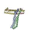
Mass: 18.015 Da / Num. of mol.: 53 / Source method: isolated from a natural source / Formula: H2O

-
Experimental details

-
Experiment

ExperimentMethod: X-RAY DIFFRACTION / Number of used crystals: 1

-
Sample preparation

CrystalDensity Matthews: 2.14 Å3/Da / Density % sol: 42.61 %
Crystal growTemperature: 293 K / Method: vapor diffusion, hanging drop / pH: 6.6
Details: 35% PEG 400, 0.1M Bis-Tris pH 6.6, VAPOR DIFFUSION, HANGING DROP, temperature 293K

-
Data collection

DiffractionMean temperature: 95 K
Diffraction sourceSource: SYNCHROTRON / Site: ALS / Beamline: 4.2.2 / Wavelength: 0.96410908, 0.99505962
DetectorType: NOIR-1 / Detector: CCD / Date: Feb 10, 2007 / Details: beamline
RadiationProtocol: SINGLE WAVELENGTH / Monochromatic (M) / Laue (L): M / Scattering type: x-ray
Radiation wavelength
IDWavelength (Å)Relative weight
10.964109081
20.995059621
ReflectionRedundancy: 17.5 % / Av σ(I) over netI: 8.7 / Number: 208914 / Rmerge(I) obs: 0.1 / Χ2: 1.33 / D res high: 2.4 Å / D res low: 30 Å / Num. obs: 11934 / % possible obs: 84.5
Diffraction reflection shell

ID: 1

Highest resolution (Å)Lowest resolution (Å)% possible obs (%)Rmerge(I) obsChi squaredRedundancyRejects
5.16301000.0642.90120.4120
4.15.1699.90.0791.7920.965
3.584.199.90.0981.4621.141
3.263.5899.90.1251.08319.935
3.023.2696.50.160.8471835
2.853.0286.20.1930.75116.630
2.72.8578.90.2390.61415.252
2.592.769.90.2520.55113.261
2.492.5958.20.2760.49411.837
2.42.4953.30.3290.4859.638
ReflectionResolution: 2.55→52.34 Å / Num. all: 11539 / Num. obs: 11398 / % possible obs: 98.8 % / Observed criterion σ(I): 5 / Redundancy: 5.96 % / Rmerge(I) obs: 0.059 / Χ2: 0.98 / Net I/σ(I): 14.5 / Scaling rejects: 514
Reflection shellResolution: 2.55→2.64 Å / Redundancy: 5.92 % / Rmerge(I) obs: 0.242 / Mean I/σ(I) obs: 5.7 / Num. measured all: 6731 / Num. unique all: 1131 / Χ2: 1.17 / % possible all: 99.9

-
Phasing

PhasingMethod: SAD

-
Processing

Software
NameVersionClassificationNB
d*TREKdata scaling
CNS1.1refinement
PDB_EXTRACT3.005data extraction
d*TREKdata reduction
PHENIXphasing
RefinementMethod to determine structure: SAD / Resolution: 2.55→39.69 Å / Rfactor Rfree error: 0.012 / Data cutoff high absF: 453570.875 / Data cutoff low absF: 0 / Isotropic thermal model: RESTRAINED / Cross valid method: THROUGHOUT / σ(F): 0 / Stereochemistry target values: Engh & Huber
RfactorNum. reflection% reflectionSelection details
Rfree0.284 583 5.2 %RANDOM
Rwork0.253 ---
all-11549 --
obs-11295 97.8 %-
Solvent computationSolvent model: FLAT MODEL / Bsol: 52.615 Å2 / ksol: 0.383 e/Å3
Displacement parametersBiso mean: 54.6 Å2
Baniso -1Baniso -2Baniso -3
1--6.28 Å22.82 Å20 Å2
2---6.28 Å20 Å2
3---12.56 Å2
Refine analyze
FreeObs
Luzzati coordinate error0.45 Å0.37 Å
Luzzati d res low-5 Å
Luzzati sigma a0.3 Å0.26 Å
Refinement stepCycle: LAST / Resolution: 2.55→39.69 Å
ProteinNucleic acidLigandSolventTotal
Num. atoms2430 0 0 53 2483
Refine LS restraints
Refine-IDTypeDev ideal
X-RAY DIFFRACTIONc_bond_d0.007
X-RAY DIFFRACTIONc_angle_deg1.2
X-RAY DIFFRACTIONc_dihedral_angle_d16.3
X-RAY DIFFRACTIONc_improper_angle_d0.7
X-RAY DIFFRACTIONc_mcbond_it
X-RAY DIFFRACTIONc_mcangle_it
X-RAY DIFFRACTIONc_scbond_it
X-RAY DIFFRACTIONc_scangle_it
LS refinement shellResolution: 2.55→2.64 Å / Rfactor Rfree error: 0.048 / Total num. of bins used: 10
RfactorNum. reflection% reflection
Rfree0.37 60 5.4 %
Rwork0.266 1056 -
all-1116 -
obs-1112 98.8 %
Xplor file
Refine-IDSerial noParam fileTopol file
X-RAY DIFFRACTION1protein_rep.paramprotein.top
X-RAY DIFFRACTION2water_rep.paramwater.top

+
About Yorodumi

-
News

-
Feb 9, 2022. New format data for meta-information of EMDB entries

New format data for meta-information of EMDB entries

  • Version 3 of the EMDB header file is now the official format.
  • The previous official version 1.9 will be removed from the archive.

Related info.:EMDB header

External links:wwPDB to switch to version 3 of the EMDB data model

-
Aug 12, 2020. Covid-19 info

Covid-19 info

URL: https://pdbjlvh1.pdbj.org/emnavi/covid19.php

New page: Covid-19 featured information page in EM Navigator.

Related info.:Covid-19 info / Mar 5, 2020. Novel coronavirus structure data

+
Mar 5, 2020. Novel coronavirus structure data

Novel coronavirus structure data

Related info.:Yorodumi Speices / Aug 12, 2020. Covid-19 info

External links:COVID-19 featured content - PDBj / Molecule of the Month (242):Coronavirus Proteases

+
Jan 31, 2019. EMDB accession codes are about to change! (news from PDBe EMDB page)

EMDB accession codes are about to change! (news from PDBe EMDB page)

  • The allocation of 4 digits for EMDB accession codes will soon come to an end. Whilst these codes will remain in use, new EMDB accession codes will include an additional digit and will expand incrementally as the available range of codes is exhausted. The current 4-digit format prefixed with “EMD-” (i.e. EMD-XXXX) will advance to a 5-digit format (i.e. EMD-XXXXX), and so on. It is currently estimated that the 4-digit codes will be depleted around Spring 2019, at which point the 5-digit format will come into force.
  • The EM Navigator/Yorodumi systems omit the EMD- prefix.

Related info.:Q: What is EMD? / ID/Accession-code notation in Yorodumi/EM Navigator

External links:EMDB Accession Codes are Changing Soon! / Contact to PDBj

+
Jul 12, 2017. Major update of PDB

Major update of PDB

  • wwPDB released updated PDB data conforming to the new PDBx/mmCIF dictionary.
  • This is a major update changing the version number from 4 to 5, and with Remediation, in which all the entries are updated.
  • In this update, many items about electron microscopy experimental information are reorganized (e.g. em_software).
  • Now, EM Navigator and Yorodumi are based on the updated data.

External links:wwPDB Remediation / Enriched Model Files Conforming to OneDep Data Standards Now Available in the PDB FTP Archive

-
Yorodumi

Thousand views of thousand structures

  • Yorodumi is a browser for structure data from EMDB, PDB, SASBDB, etc.
  • This page is also the successor to EM Navigator detail page, and also detail information page/front-end page for Omokage search.
  • The word "yorodu" (or yorozu) is an old Japanese word meaning "ten thousand". "mi" (miru) is to see.

Related info.:EMDB / PDB / SASBDB / Comparison of 3 databanks / Yorodumi Search / Aug 31, 2016. New EM Navigator & Yorodumi / Yorodumi Papers / Jmol/JSmol / Function and homology information / Changes in new EM Navigator and Yorodumi

Read more