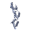
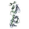
regulation of spindle assembly / regulation of kinetochore assembly / SUMO polymer binding / response to arsenic-containing substance / protein K11-linked ubiquitination / protein K6-linked ubiquitination / protein K63-linked ubiquitination / nucleosome binding / protein autoubiquitination / negative regulation of protein localization to chromatin ...regulation of spindle assembly / regulation of kinetochore assembly / SUMO polymer binding / response to arsenic-containing substance / protein K11-linked ubiquitination / protein K6-linked ubiquitination / protein K63-linked ubiquitination / nucleosome binding / protein autoubiquitination / negative regulation of protein localization to chromatin / protein K48-linked ubiquitination / PML body / RING-type E3 ubiquitin transferase / ubiquitin-protein transferase activity / ubiquitin protein ligase activity / Antigen processing: Ubiquitination & Proteasome degradation / Processing of DNA double-strand break ends / proteasome-mediated ubiquitin-dependent protein catabolic process / nuclear body / positive regulation of DNA-templated transcription / DNA-templated transcription / positive regulation of transcription by RNA polymerase II / DNA binding / zinc ion binding / nucleoplasm / identical protein binding / nucleus / cytoplasm Similarity search - Function
RNF4, RING finger, HC subclass / : / Zinc finger, C3HC4 type (RING finger) / Ring finger domain / Zinc/RING finger domain, C3HC4 (zinc finger) / Herpes Virus-1 / Zinc finger, RING-type, conserved site / Zinc finger RING-type signature. / Ring finger / Zinc finger RING-type profile. ...RNF4, RING finger, HC subclass / : / Zinc finger, C3HC4 type (RING finger) / Ring finger domain / Zinc/RING finger domain, C3HC4 (zinc finger) / Herpes Virus-1 / Zinc finger, RING-type, conserved site / Zinc finger RING-type signature. / Ring finger / Zinc finger RING-type profile. / Zinc finger, RING-type / Zinc finger, RING/FYVE/PHD-type / 2-Layer Sandwich / Alpha Beta Similarity search - Domain/homology
In the structure databanks used in Yorodumi, some data are registered as the other names, "COVID-19 virus" and "2019-nCoV". Here are the details of the virus and the list of structure data.
Jan 31, 2019. EMDB accession codes are about to change! (news from PDBe EMDB page)
EMDB accession codes are about to change! (news from PDBe EMDB page)
The allocation of 4 digits for EMDB accession codes will soon come to an end. Whilst these codes will remain in use, new EMDB accession codes will include an additional digit and will expand incrementally as the available range of codes is exhausted. The current 4-digit format prefixed with “EMD-” (i.e. EMD-XXXX) will advance to a 5-digit format (i.e. EMD-XXXXX), and so on. It is currently estimated that the 4-digit codes will be depleted around Spring 2019, at which point the 5-digit format will come into force.
The EM Navigator/Yorodumi systems omit the EMD- prefix.
Related info.:Q: What is EMD? / ID/Accession-code notation in Yorodumi/EM Navigator
Yorodumi is a browser for structure data from EMDB, PDB, SASBDB, etc.
This page is also the successor to EM Navigator detail page, and also detail information page/front-end page for Omokage search.
The word "yorodu" (or yorozu) is an old Japanese word meaning "ten thousand". "mi" (miru) is to see.
Related info.:EMDB / PDB / SASBDB / Comparison of 3 databanks / Yorodumi Search / Aug 31, 2016. New EM Navigator & Yorodumi / Yorodumi Papers / Jmol/JSmol / Function and homology information / Changes in new EM Navigator and Yorodumi
 Movie
Movie Controller


 Open data
 Basic information
 Components
 Keywords
 Function and homology information
 Homo sapiens (human)
 X-RAY DIFFRACTION /
  Authors
 Citation
 Structure visualization
 Downloads & links
 Other downloads


 PDBj
PDBj

 Assembly
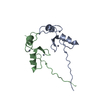


 Mass: 65.409 Da / Num. of mol.: 4 / Source method: obtained synthetically / Formula: Zn
 Mass: 18.015 Da / Num. of mol.: 70 / Source method: isolated from a natural source / Formula: H2O
 Sample preparation
 / Beamline: 12.3.1 / Wavelength: 1 Å
 Processing