Protocol: SINGLE WAVELENGTH / Monochromatic (M) / Laue (L): M / Scattering type: x-ray
Radiation wavelength
Wavelength: 0.9796 Å / Relative weight: 1
Reflection
Resolution: 2.9→67.49 Å / Num. obs: 2864 / % possible obs: 100 % / Observed criterion σ(I): 2 / Redundancy: 13.4 % / Rmerge(I) obs: 0.13 / Net I/σ(I): 13.3
Reflection shell
Resolution: 2.9→3.06 Å / Redundancy: 14.4 % / Rmerge(I) obs: 1.25 / Mean I/σ(I) obs: 3.1 / % possible all: 100
-
Processing
Software
Name
Version
Classification
PHENIX
(PHENIX.REFINE)
refinement
MOSFLM
datareduction
SCALA
datascaling
SCALA
phasing
Refinement
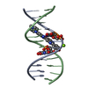
Method to determine structure: SAD Starting model: NONE Resolution: 2.901→47.723 Å / SU ML: 0.41 / σ(F): 1.38 / Phase error: 37.35 / Stereochemistry target values: ML Details: ONLY RESIDUES 16-44 ARE VISIBLE IN THE ELECTION DENSITY. RESIDUES 6-15 AND 45-64 WERE NOT MODELLED.
Rfactor
Num. reflection
% reflection
Rfree
0.2915
228
4.6 %
Rwork
0.2675
-
-
obs
0.2687
4949
99.98 %
Solvent computation
Shrinkage radii: 0.9 Å / VDW probe radii: 1.11 Å / Solvent model: FLAT BULK SOLVENT MODEL
Refinement step
Cycle: LAST / Resolution: 2.901→47.723 Å
Protein
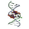
Nucleic acid
Ligand
Solvent
Total
Num. atoms
641
0
0
0
641
Refine LS restraints
Refine-ID
Type
Dev ideal
Number
X-RAY DIFFRACTION
f_bond_d
0.006
651
X-RAY DIFFRACTION
f_angle_d
1.02
875
X-RAY DIFFRACTION
f_dihedral_angle_d
23.604
259
X-RAY DIFFRACTION
f_chiral_restr
0.052
106
X-RAY DIFFRACTION
f_plane_restr
0.005
119
LS refinement shell
Resolution (Å)
Rfactor Rfree
Num. reflection Rfree
Rfactor Rwork
Num. reflection Rwork
Refine-ID
% reflection obs (%)
2.9014-3.6552
0.3861
108
0.3012
2366
X-RAY DIFFRACTION
100
3.6552-47.7299
0.2613
120
0.2558
2355
X-RAY DIFFRACTION
100
+
About Yorodumi
-
News
-
Feb 9, 2022. New format data for meta-information of EMDB entries
New format data for meta-information of EMDB entries
Version 3 of the EMDB header file is now the official format.
The previous official version 1.9 will be removed from the archive.
In the structure databanks used in Yorodumi, some data are registered as the other names, "COVID-19 virus" and "2019-nCoV". Here are the details of the virus and the list of structure data.
Jan 31, 2019. EMDB accession codes are about to change! (news from PDBe EMDB page)
EMDB accession codes are about to change! (news from PDBe EMDB page)
The allocation of 4 digits for EMDB accession codes will soon come to an end. Whilst these codes will remain in use, new EMDB accession codes will include an additional digit and will expand incrementally as the available range of codes is exhausted. The current 4-digit format prefixed with “EMD-” (i.e. EMD-XXXX) will advance to a 5-digit format (i.e. EMD-XXXXX), and so on. It is currently estimated that the 4-digit codes will be depleted around Spring 2019, at which point the 5-digit format will come into force.
The EM Navigator/Yorodumi systems omit the EMD- prefix.
Related info.:Q: What is EMD? / ID/Accession-code notation in Yorodumi/EM Navigator
Yorodumi is a browser for structure data from EMDB, PDB, SASBDB, etc.
This page is also the successor to EM Navigator detail page, and also detail information page/front-end page for Omokage search.
The word "yorodu" (or yorozu) is an old Japanese word meaning "ten thousand". "mi" (miru) is to see.
Related info.:EMDB / PDB / SASBDB / Comparison of 3 databanks / Yorodumi Search / Aug 31, 2016. New EM Navigator & Yorodumi / Yorodumi Papers / Jmol/JSmol / Function and homology information / Changes in new EM Navigator and Yorodumi
 Movie
Movie Controller


 Open data
 Basic information
 Components
 Keywords
 Function and homology information
 HOMO SAPIENS (human)
 X-RAY DIFFRACTION /
  Authors
 Citation
 Structure visualization
 Downloads & links
 Other downloads
 PDBj
PDBj
 Assembly

 Sample preparation
 / Beamline: I02 / Wavelength: 0.9796
  Processing