[English] 日本語
Yorodumi
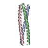
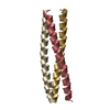
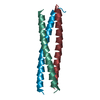
- PDB-3vyi: Crystal Structure of a trimeric coiled-coil (I/I-type) assembly d... -

+
Open data


ID or keywords:

Loading...

-
Basic information

Entry
Database: PDB / ID: 3vyi
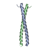
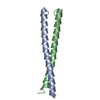
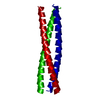
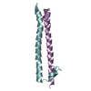
TitleCrystal Structure of a trimeric coiled-coil (I/I-type) assembly domain from the voltage-gated proton channel mutant
ComponentsVoltage-gated hydrogen channel 1
KeywordsMEMBRANE PROTEIN / coiled-coil / assembly / cytoplasmic
Function / homology
Function and homology information


voltage-gated proton channel activity / Sperm Motility And Taxes / regulation of acrosome reaction / ROS and RNS production in phagocytes / cellular response to pH / regulation of reactive oxygen species biosynthetic process / response to pH / cellular response to zinc ion / monoatomic ion channel complex / response to zinc ion ...voltage-gated proton channel activity / Sperm Motility And Taxes / regulation of acrosome reaction / ROS and RNS production in phagocytes / cellular response to pH / regulation of reactive oxygen species biosynthetic process / response to pH / cellular response to zinc ion / monoatomic ion channel complex / response to zinc ion / sperm flagellum / proton transmembrane transport / Neutrophil degranulation / positive regulation of superoxide anion generation / regulation of intracellular pH / phagocytic vesicle membrane / apical plasma membrane / innate immune response / protein homodimerization activity / membrane / identical protein binding / plasma membrane
Similarity search - Function
Voltage-gated hydrogen channel 1, C-terminal membrane-localisation domain / Voltage-gated hydrogen channel 1 / C-terminal membrane-localisation domain of ion-channel, VCN1 / Single alpha-helices involved in coiled-coils or other helix-helix interfaces - #170 / Voltage-dependent channel domain superfamily / Single alpha-helices involved in coiled-coils or other helix-helix interfaces / Ion transport domain / Ion transport protein / Up-down Bundle / Mainly Alpha
Similarity search - Domain/homology
Voltage-gated hydrogen channel 1
Similarity search - Component
Biological speciesMus musculus (house mouse)
MethodX-RAY DIFFRACTION / SYNCHROTRON / MOLECULAR REPLACEMENT / Resolution: 2.305 Å
AuthorsFujiwara, Y. / Takeshita, K. / Nakagawa, A.
CitationJournal: J.Physiol.(Paris) / Year: 2013
Title: Gating of the designed trimeric/tetrameric voltage-gated H+ channel.
Authors: Fujiwara, Y. / Kurokawa, T. / Takeshita, K. / Nakagawa, A. / Larsson, H.P. / Okamura, Y.
History
DepositionSep 25, 2012Deposition site: PDBJ / Processing site: PDBJ
Revision 1.0Feb 27, 2013Provider: repository / Type: Initial release
Revision 1.1Mar 20, 2024Group: Data collection / Database references
Category: chem_comp_atom / chem_comp_bond ...chem_comp_atom / chem_comp_bond / database_2 / struct_ref_seq_dif
Item: _database_2.pdbx_DOI / _database_2.pdbx_database_accession / _struct_ref_seq_dif.details

-
Structure visualization

Structure viewerMolecule:
MolmilJmol/JSmol

Downloads & links

-
Assembly

Deposited unit
A: Voltage-gated hydrogen channel 1
B: Voltage-gated hydrogen channel 1
D: Voltage-gated hydrogen channel 1
C: Voltage-gated hydrogen channel 1
E: Voltage-gated hydrogen channel 1
F: Voltage-gated hydrogen channel 1
H: Voltage-gated hydrogen channel 1
G: Voltage-gated hydrogen channel 1
I: Voltage-gated hydrogen channel 1
J: Voltage-gated hydrogen channel 1
K: Voltage-gated hydrogen channel 1
L: Voltage-gated hydrogen channel 1


Theoretical massNumber of molelcules
Total (without water)71,06112
Polymers71,06112
Non-polymers00
Water2,882160
1
A: Voltage-gated hydrogen channel 1
B: Voltage-gated hydrogen channel 1
C: Voltage-gated hydrogen channel 1


Theoretical massNumber of molelcules
Total (without water)17,7653
Polymers17,7653
Non-polymers00
Water543
TypeNameSymmetry operationNumber
identity operation1_555x,y,z1
Buried area4990 Å2
ΔGint-52 kcal/mol
Surface area9320 Å2
MethodPISA
2
D: Voltage-gated hydrogen channel 1
E: Voltage-gated hydrogen channel 1
F: Voltage-gated hydrogen channel 1


Theoretical massNumber of molelcules
Total (without water)17,7653
Polymers17,7653
Non-polymers00
Water543
TypeNameSymmetry operationNumber
identity operation1_555x,y,z1
Buried area4880 Å2
ΔGint-51 kcal/mol
Surface area9450 Å2
MethodPISA
3
H: Voltage-gated hydrogen channel 1
G: Voltage-gated hydrogen channel 1
I: Voltage-gated hydrogen channel 1


Theoretical massNumber of molelcules
Total (without water)17,7653
Polymers17,7653
Non-polymers00
Water543
TypeNameSymmetry operationNumber
identity operation1_555x,y,z1
Buried area4130 Å2
ΔGint-45 kcal/mol
Surface area8450 Å2
MethodPISA
4
J: Voltage-gated hydrogen channel 1
K: Voltage-gated hydrogen channel 1
L: Voltage-gated hydrogen channel 1


Theoretical massNumber of molelcules
Total (without water)17,7653
Polymers17,7653
Non-polymers00
Water543
TypeNameSymmetry operationNumber
identity operation1_555x,y,z1
Buried area4220 Å2
ΔGint-44 kcal/mol
Surface area8440 Å2
MethodPISA
Unit cell
Length a, b, c (Å)83.273, 84.010, 88.393
Angle α, β, γ (deg.)90.00, 90.00, 90.00
Int Tables number19
Space group name H-MP212121

-
Components

#1: Protein
Voltage-gated hydrogen channel 1 / Hydrogen voltage-gated channel 1 / HV1 / Voltage sensor domain-only protein / mVSOP


Mass: 5921.762 Da / Num. of mol.: 12 / Fragment: C-TERMINAL DOMAIN, UNP RESIDUES 220-269
Mutation: L227I, I230N, N231I, I232N, L234L, L241I, C245I, K248I, l255I, L259I
Source method: isolated from a genetically manipulated source
Source: (gene. exp.) Mus musculus (house mouse) / Gene: Hvcn1, Bts, Vsop / Production host: Escherichia coli (E. coli) / References: UniProt: Q3U2S8
#2: Water ChemComp-HOH / water


Mass: 18.015 Da / Num. of mol.: 160 / Source method: isolated from a natural source / Formula: H2O

-
Experimental details

-
Experiment

ExperimentMethod: X-RAY DIFFRACTION / Number of used crystals: 1

-
Sample preparation

CrystalDensity Matthews: 2.18 Å3/Da / Density % sol: 43.46 %
Crystal growTemperature: 293 K / Method: vapor diffusion, hanging drop / pH: 8.5
Details: 0.2M AmSO4, 0.1M Tris-HCl (pH 8.5), 25% w/v PEG 3350, VAPOR DIFFUSION, HANGING DROP, temperature 293K

-
Data collection

DiffractionMean temperature: 77 K
Diffraction sourceSource: SYNCHROTRON / Site: SPring-8 / Beamline: BL44XU / Wavelength: 0.9 Å
DetectorType: RAYONIX MX225HE / Detector: CCD / Date: Jul 22, 2011
RadiationProtocol: SINGLE WAVELENGTH / Monochromatic (M) / Laue (L): M / Scattering type: x-ray
Radiation wavelengthWavelength: 0.9 Å / Relative weight: 1
ReflectionResolution: 2.3→50 Å / Num. all: 27731 / Num. obs: 27673 / % possible obs: 99.1 % / Observed criterion σ(F): 1.35 / Observed criterion σ(I): 31.07
Reflection shell
Resolution (Å)Diffraction-ID% possible all
2.3-2.34197.7
2.34-2.38198.6
2.38-2.43198.7
2.43-2.48198.8
2.48-2.53199.7
2.53-2.59199.4
2.59-2.66199.5
2.66-2.73199.6
2.73-2.81199.6
2.81-2.9199.8
2.9-3199.7
3-3.12199.9
3.12-3.26199.9
3.26-3.44199.9
3.44-3.65199.9
3.65-3.93199.9
3.93-4.331100
4.33-4.951100
4.95-6.24199.9
6.24-50192.4

-
Processing

Software
NameVersionClassification
HKL-2000data collection
MOLREP& PHASERphasing
PHENIX(phenix.refine: 1.7.3_928)refinement
HKL-2000data reduction
SCALEPACKdata scaling
RefinementMethod to determine structure: MOLECULAR REPLACEMENT / Resolution: 2.305→39.114 Å / SU ML: 0.3 / σ(F): 1.35 / Phase error: 31.1 / Stereochemistry target values: ML
RfactorNum. reflection% reflection
Rfree0.2948 1396 5.04 %
Rwork0.2324 --
obs0.2356 27673 98.74 %
all-27731 -
Solvent computationShrinkage radii: 0.9 Å / VDW probe radii: 1.11 Å / Solvent model: FLAT BULK SOLVENT MODEL / Bsol: 46.401 Å2 / ksol: 0.363 e/Å3
Displacement parameters
Baniso -1Baniso -2Baniso -3
1--6.6009 Å20 Å20 Å2
2---0.1526 Å2-0 Å2
3---6.7535 Å2
Refinement stepCycle: LAST / Resolution: 2.305→39.114 Å
ProteinNucleic acidLigandSolventTotal
Num. atoms4369 0 0 160 4529
Refine LS restraints
Refine-IDTypeDev idealNumber
X-RAY DIFFRACTIONf_bond_d0.0094381
X-RAY DIFFRACTIONf_angle_d1.0265835
X-RAY DIFFRACTIONf_dihedral_angle_d18.9381799
X-RAY DIFFRACTIONf_chiral_restr0.069690
X-RAY DIFFRACTIONf_plane_restr0.004752
LS refinement shell

Refine-ID: X-RAY DIFFRACTION / Total num. of bins used: 10

Resolution (Å)Rfactor RfreeNum. reflection RfreeRfactor RworkNum. reflection Rwork% reflection obs (%)
2.3046-2.38690.33691300.244247494
2.3869-2.48250.2721380.228257699
2.4825-2.59540.33891400.21652629100
2.5954-2.73220.28881380.2303258899
2.7322-2.90340.30981390.23962654100
2.9034-3.12740.33011270.24542646100
3.1274-3.4420.27391310.24172648100
3.442-3.93970.27441680.21442651100
3.9397-4.9620.2681340.20292705100
4.962-39.11960.32541510.2725270696

+
About Yorodumi

-
News

-
Feb 9, 2022. New format data for meta-information of EMDB entries

New format data for meta-information of EMDB entries

  • Version 3 of the EMDB header file is now the official format.
  • The previous official version 1.9 will be removed from the archive.

Related info.:EMDB header

External links:wwPDB to switch to version 3 of the EMDB data model

-
Aug 12, 2020. Covid-19 info

Covid-19 info

URL: https://pdbjlvh1.pdbj.org/emnavi/covid19.php

New page: Covid-19 featured information page in EM Navigator.

Related info.:Covid-19 info / Mar 5, 2020. Novel coronavirus structure data

+
Mar 5, 2020. Novel coronavirus structure data

Novel coronavirus structure data

Related info.:Yorodumi Speices / Aug 12, 2020. Covid-19 info

External links:COVID-19 featured content - PDBj / Molecule of the Month (242):Coronavirus Proteases

+
Jan 31, 2019. EMDB accession codes are about to change! (news from PDBe EMDB page)

EMDB accession codes are about to change! (news from PDBe EMDB page)

  • The allocation of 4 digits for EMDB accession codes will soon come to an end. Whilst these codes will remain in use, new EMDB accession codes will include an additional digit and will expand incrementally as the available range of codes is exhausted. The current 4-digit format prefixed with “EMD-” (i.e. EMD-XXXX) will advance to a 5-digit format (i.e. EMD-XXXXX), and so on. It is currently estimated that the 4-digit codes will be depleted around Spring 2019, at which point the 5-digit format will come into force.
  • The EM Navigator/Yorodumi systems omit the EMD- prefix.

Related info.:Q: What is EMD? / ID/Accession-code notation in Yorodumi/EM Navigator

External links:EMDB Accession Codes are Changing Soon! / Contact to PDBj

+
Jul 12, 2017. Major update of PDB

Major update of PDB

  • wwPDB released updated PDB data conforming to the new PDBx/mmCIF dictionary.
  • This is a major update changing the version number from 4 to 5, and with Remediation, in which all the entries are updated.
  • In this update, many items about electron microscopy experimental information are reorganized (e.g. em_software).
  • Now, EM Navigator and Yorodumi are based on the updated data.

External links:wwPDB Remediation / Enriched Model Files Conforming to OneDep Data Standards Now Available in the PDB FTP Archive

-
Yorodumi

Thousand views of thousand structures

  • Yorodumi is a browser for structure data from EMDB, PDB, SASBDB, etc.
  • This page is also the successor to EM Navigator detail page, and also detail information page/front-end page for Omokage search.
  • The word "yorodu" (or yorozu) is an old Japanese word meaning "ten thousand". "mi" (miru) is to see.

Related info.:EMDB / PDB / SASBDB / Comparison of 3 databanks / Yorodumi Search / Aug 31, 2016. New EM Navigator & Yorodumi / Yorodumi Papers / Jmol/JSmol / Function and homology information / Changes in new EM Navigator and Yorodumi

Read more