- PDB-3vmz: Crystal Structure of a parallel coiled-coil dimerization domain f... -
+
Open data
ID or keywords:
Loading...
-
Basic information
Entry
Database: PDB / ID: 3vmz
Title
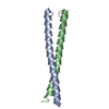
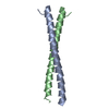
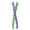
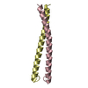
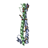
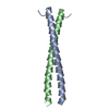
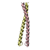
Crystal Structure of a parallel coiled-coil dimerization domain from the voltage-gated proton channel (oxidation/H2O2)
Components
Voltage-gated hydrogen channel 1
Keywords
MEMBRANE PROTEIN / COILED-COIL / ASSEMBLY DOMAIN / ION TRANSPORT
Function / homology
Function and homology information
voltage-gated proton channel activity / Sperm Motility And Taxes / regulation of acrosome reaction / ROS and RNS production in phagocytes / cellular response to pH / response to pH / regulation of reactive oxygen species biosynthetic process / voltage-gated monoatomic cation channel activity / response to zinc ion / cellular response to zinc ion ...voltage-gated proton channel activity / Sperm Motility And Taxes / regulation of acrosome reaction / ROS and RNS production in phagocytes / cellular response to pH / response to pH / regulation of reactive oxygen species biosynthetic process / voltage-gated monoatomic cation channel activity / response to zinc ion / cellular response to zinc ion / monoatomic ion channel complex / sperm flagellum / positive regulation of superoxide anion generation / Neutrophil degranulation / proton transmembrane transport / regulation of intracellular pH / phagocytic vesicle membrane / apical plasma membrane / innate immune response / protein homodimerization activity / membrane / identical protein binding / plasma membrane Similarity search - Function
Voltage-gated hydrogen channel 1, C-terminal membrane-localisation domain / Voltage-gated hydrogen channel 1 / C-terminal membrane-localisation domain of ion-channel, VCN1 / Single alpha-helices involved in coiled-coils or other helix-helix interfaces - #170 / Voltage-dependent channel domain superfamily / Single alpha-helices involved in coiled-coils or other helix-helix interfaces / Ion transport domain / Ion transport protein / Up-down Bundle / Mainly Alpha Similarity search - Domain/homology
Resolution: 1.55→40.94 Å / Cor.coef. Fo:Fc: 0.96 / Cor.coef. Fo:Fc free: 0.938 / SU B: 3.762 / SU ML: 0.062 / Cross valid method: THROUGHOUT / σ(F): 0 / ESU R Free: 0.1 / Stereochemistry target values: MAXIMUM LIKELIHOOD Details: HYDROGENS HAVE BEEN ADDED IN THE RIDING POSITIONS U VALUES
Rfactor
Num. reflection
% reflection
Selection details
Rfree
0.233
1346
5 %
RANDOM
Rwork
0.193
-
-
-
obs
0.195
26774
99.7 %
-
Solvent computation
Ion probe radii: 0.8 Å / Shrinkage radii: 0.8 Å / VDW probe radii: 1.4 Å / Solvent model: MASK
Displacement parameters
Biso mean: 23.77 Å2
Baniso -1
Baniso -2
Baniso -3
1-
0.09 Å2
0 Å2
0 Å2
2-
-
0.08 Å2
0 Å2
3-
-
-
-0.17 Å2
Refinement step
Cycle: LAST / Resolution: 1.55→40.94 Å
Protein
Nucleic acid
Ligand
Solvent
Total
Num. atoms
1556
0
0
225
1781
Refine LS restraints
Refine-ID
Type
Dev ideal
Dev ideal target
Number
X-RAY DIFFRACTION
r_bond_refined_d
0.025
0.022
1660
X-RAY DIFFRACTION
r_bond_other_d
X-RAY DIFFRACTION
r_angle_refined_deg
2.36
1.997
2203
X-RAY DIFFRACTION
r_angle_other_deg
X-RAY DIFFRACTION
r_dihedral_angle_1_deg
4.142
5
187
X-RAY DIFFRACTION
r_dihedral_angle_2_deg
42.799
26.222
90
X-RAY DIFFRACTION
r_dihedral_angle_3_deg
15.855
15
417
X-RAY DIFFRACTION
r_dihedral_angle_4_deg
19.931
15
13
X-RAY DIFFRACTION
r_chiral_restr
0.175
0.2
261
X-RAY DIFFRACTION
r_gen_planes_refined
0.01
0.02
1141
X-RAY DIFFRACTION
r_gen_planes_other
X-RAY DIFFRACTION
r_nbd_refined
X-RAY DIFFRACTION
r_nbd_other
X-RAY DIFFRACTION
r_nbtor_refined
X-RAY DIFFRACTION
r_nbtor_other
X-RAY DIFFRACTION
r_xyhbond_nbd_refined
X-RAY DIFFRACTION
r_xyhbond_nbd_other
X-RAY DIFFRACTION
r_metal_ion_refined
X-RAY DIFFRACTION
r_metal_ion_other
X-RAY DIFFRACTION
r_symmetry_vdw_refined
X-RAY DIFFRACTION
r_symmetry_vdw_other
X-RAY DIFFRACTION
r_symmetry_hbond_refined
X-RAY DIFFRACTION
r_symmetry_hbond_other
X-RAY DIFFRACTION
r_symmetry_metal_ion_refined
X-RAY DIFFRACTION
r_symmetry_metal_ion_other
X-RAY DIFFRACTION
r_mcbond_it
1.512
1.5
960
X-RAY DIFFRACTION
r_mcbond_other
X-RAY DIFFRACTION
r_mcangle_it
2.404
2
1567
X-RAY DIFFRACTION
r_scbond_it
3.749
3
700
X-RAY DIFFRACTION
r_scangle_it
6.164
4.5
636
X-RAY DIFFRACTION
r_rigid_bond_restr
X-RAY DIFFRACTION
r_sphericity_free
X-RAY DIFFRACTION
r_sphericity_bonded
LS refinement shell
Resolution: 1.55→1.59 Å / Total num. of bins used: 20
Rfactor
Num. reflection
% reflection
Rfree
0.269
96
-
Rwork
0.231
1774
-
obs
-
-
97.65 %
Refinement TLS params.
Method: refined / Refine-ID: X-RAY DIFFRACTION
ID
L11 (°2)
L12 (°2)
L13 (°2)
L22 (°2)
L23 (°2)
L33 (°2)
S11 (Å °)
S12 (Å °)
S13 (Å °)
S21 (Å °)
S22 (Å °)
S23 (Å °)
S31 (Å °)
S32 (Å °)
S33 (Å °)
T11 (Å2)
T12 (Å2)
T13 (Å2)
T22 (Å2)
T23 (Å2)
T33 (Å2)
Origin x (Å)
Origin y (Å)
Origin z (Å)
1
0.543
0.5229
0.0102
2.5763
-1.4277
1.1248
0.0255
-0.0913
-0.0776
-0.0721
-0.0422
-0.015
0.0404
-0.0079
0.0166
0.0572
0.0148
0.0169
0.0497
0.039
0.0683
33.0991
32.584
17.0078
2
0.8273
-1.0022
-0.149
1.7375
0.101
0.1213
0.0693
0.0051
0.0971
0.0541
-0.0838
-0.1311
-0.052
0.0621
0.0145
0.0861
-0.0038
0.0176
0.107
-0.0088
0.0748
45.5096
31.9231
26.3005
Refinement TLS group
ID
Refine-ID
Refine TLS-ID
Auth asym-ID
Auth seq-ID
1
X-RAY DIFFRACTION
1
A
220 - 265
2
X-RAY DIFFRACTION
1
B
220 - 265
3
X-RAY DIFFRACTION
2
C
220 - 265
4
X-RAY DIFFRACTION
2
D
220 - 265
+
About Yorodumi
-
News
-
Feb 9, 2022. New format data for meta-information of EMDB entries
New format data for meta-information of EMDB entries
Version 3 of the EMDB header file is now the official format.
The previous official version 1.9 will be removed from the archive.
In the structure databanks used in Yorodumi, some data are registered as the other names, "COVID-19 virus" and "2019-nCoV". Here are the details of the virus and the list of structure data.
Jan 31, 2019. EMDB accession codes are about to change! (news from PDBe EMDB page)
EMDB accession codes are about to change! (news from PDBe EMDB page)
The allocation of 4 digits for EMDB accession codes will soon come to an end. Whilst these codes will remain in use, new EMDB accession codes will include an additional digit and will expand incrementally as the available range of codes is exhausted. The current 4-digit format prefixed with “EMD-” (i.e. EMD-XXXX) will advance to a 5-digit format (i.e. EMD-XXXXX), and so on. It is currently estimated that the 4-digit codes will be depleted around Spring 2019, at which point the 5-digit format will come into force.
The EM Navigator/Yorodumi systems omit the EMD- prefix.
Related info.:Q: What is EMD? / ID/Accession-code notation in Yorodumi/EM Navigator
Yorodumi is a browser for structure data from EMDB, PDB, SASBDB, etc.
This page is also the successor to EM Navigator detail page, and also detail information page/front-end page for Omokage search.
The word "yorodu" (or yorozu) is an old Japanese word meaning "ten thousand". "mi" (miru) is to see.
Related info.:EMDB / PDB / SASBDB / Comparison of 3 databanks / Yorodumi Search / Aug 31, 2016. New EM Navigator & Yorodumi / Yorodumi Papers / Jmol/JSmol / Function and homology information / Changes in new EM Navigator and Yorodumi
 Movie
Movie Controller

 Yorodumi
 Open data
 Basic information
 Components
 Keywords
 Function and homology information
 X-RAY DIFFRACTION /
  Authors
 Citation
 Structure visualization
 Downloads & links
 Other downloads


 PDBj
PDBj


 Assembly
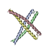

 Mass: 18.015 Da / Num. of mol.: 225 / Source method: isolated from a natural source / Formula: H2O
 Sample preparation
 / Beamline: BL44XU / Wavelength: 0.9
  Processing