 Movie
Movie Controller


+ Open data

- Basic information
| Entry | Database: PDB / ID: 4l0n | ||||||
|---|---|---|---|---|---|---|---|
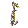
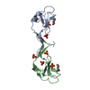
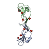
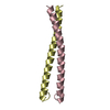
| Title | Crystal structure of STK3 (MST2) SARAH domain | ||||||
 Components Components | Serine/threonine-protein kinase 3 | ||||||
 Keywords Keywords | TRANSFERASE / Structural Genomics / Structural Genomics Consortium / SGC / SARAH domain / STK3 / MST2 / heptad repeat / structural genomics consortium (SGC) | ||||||
| Function / homology |  Function and homology information Function and homology informationnegative regulation of cell growth involved in contact inhibition / regulation of cell differentiation involved in embryonic placenta development / primitive hemopoiesis / cell differentiation involved in embryonic placenta development / positive regulation of extrinsic apoptotic signaling pathway via death domain receptors / neural tube formation / negative regulation of organ growth / positive regulation of hippo signaling / endocardium development / hippo signaling ...negative regulation of cell growth involved in contact inhibition / regulation of cell differentiation involved in embryonic placenta development / primitive hemopoiesis / cell differentiation involved in embryonic placenta development / positive regulation of extrinsic apoptotic signaling pathway via death domain receptors / neural tube formation / negative regulation of organ growth / positive regulation of hippo signaling / endocardium development / hippo signaling / organ growth / transcription regulator activator activity / Signaling by Hippo / protein localization to centrosome / hepatocyte apoptotic process / extrinsic apoptotic signaling pathway via death domain receptors / regulation of MAPK cascade / canonical Wnt signaling pathway / positive regulation of fat cell differentiation / JNK cascade / epithelial cell proliferation / protein serine/threonine kinase activator activity / central nervous system development / phosphatidylinositol 3-kinase/protein kinase B signal transduction / protein tetramerization / negative regulation of canonical Wnt signaling pathway / protein import into nucleus / positive regulation of JNK cascade / negative regulation of epithelial cell proliferation / protein phosphorylation / protein kinase activity / positive regulation of phosphatidylinositol 3-kinase/protein kinase B signal transduction / non-specific serine/threonine protein kinase / protein stabilization / intracellular signal transduction / positive regulation of apoptotic process / protein serine kinase activity / protein serine/threonine kinase activity / apoptotic process / centrosome / magnesium ion binding / protein-containing complex / ATP binding / identical protein binding / nucleus / cytosol / cytoplasm Similarity search - Function | ||||||
| Biological species |  Homo sapiens (human) Homo sapiens (human) | ||||||
| Method |  X-RAY DIFFRACTION / SYNCHROTRON / SAD / Resolution: 1.4 Å X-RAY DIFFRACTION / SYNCHROTRON / SAD / Resolution: 1.4 Å | ||||||
 Authors Authors | Chaikuad, A. / Krojer, T. / Newman, J.A. / Dixon-Clarke, S. / von Delft, F. / Arrowsmith, C.H. / Edwards, A.M. / Bountra, C. / Knapp, S. / Structural Genomics Consortium (SGC) | ||||||
 Citation Citation | Journal: To be Published Title: Crystal structure of STK3 (MST2) SARAH domain Authors: Chaikuad, A. / Krojer, T. / Newman, J.A. / Dixon-Clarke, S. / von Delft, F. / Arrowsmith, C.H. / Edwards, A.M. / Bountra, C. / Knapp, S. / Structural Genomics Consortium (SGC) | ||||||
| History |
|
- Structure visualization
| Structure viewer | Molecule: MolmilJmol/JSmol |
|---|
- Downloads & links
-Download
| PDBx/mmCIF format | 4l0n.cif.gz | 259.6 KB | Display | PDBx/mmCIF format |
|---|---|---|---|---|
| PDB format | pdb4l0n.ent.gz | 216.3 KB | Display | PDB format |
| PDBx/mmJSON format | 4l0n.json.gz | Tree view | PDBx/mmJSON format | |
| Others |  Other downloads Other downloads |
-Validation report
| Arichive directory | https://data.pdbj.org/pub/pdb/validation_reports/l0/4l0nftp://data.pdbj.org/pub/pdb/validation_reports/l0/4l0n | HTTPS FTP |
|---|
-Related structure data
| Similar structure data |
|---|


-Links
 PDBj
PDBj


- Assembly
| Deposited unit | 
| |||||||||
|---|---|---|---|---|---|---|---|---|---|---|
| 1 | 
| |||||||||
| 2 | 
| |||||||||
| 3 | 
| |||||||||
| 4 | 
| |||||||||
| 5 | 
| |||||||||
| 6 | 
| |||||||||
| Unit cell |
| |||||||||
| Components on special symmetry positions |
|
-Components
| #1: Protein | Mass: 6168.191 Da / Num. of mol.: 10 / Fragment: SARAH domain (UNP residues 436-484) Source method: isolated from a genetically manipulated source Source: (gene. exp.) Homo sapiens (human) / Gene: KRS1, MST2, STK3 / Plasmid: pNIC28-BSA4 / Production host:  References: UniProt: Q13188, non-specific serine/threonine protein kinase #2: Water | ChemComp-HOH / |  Mass: 18.015 Da / Num. of mol.: 799 / Source method: isolated from a natural source / Formula: H2O Mass: 18.015 Da / Num. of mol.: 799 / Source method: isolated from a natural source / Formula: H2O |
|---|
-Experimental details
-Experiment
| Experiment | Method: X-RAY DIFFRACTION / Number of used crystals: 1 |
|---|
- Sample preparation
| Crystal | Density Matthews: 2.3 Å3/Da / Density % sol: 46.53 % |
|---|---|
| Crystal grow | Temperature: 277.15 K / Method: vapor diffusion, sitting drop / pH: 4.9 Details: 30% MPD, 0.1M acetate pH 4.9, 0.2M calcium chloride, VAPOR DIFFUSION, SITTING DROP, temperature 277.15K |
-Data collection
| Diffraction | Mean temperature: 100 K |
|---|---|
| Diffraction source | Source: SYNCHROTRON / Site: Diamond  / Beamline: I02 / Wavelength: 0.97949 Å / Beamline: I02 / Wavelength: 0.97949 Å |
| Detector | Type: PSI PILATUS 6M / Detector: PIXEL / Date: Apr 19, 2013 |
| Radiation | Monochromator: Si (111) double crystal monochromator / Protocol: SINGLE WAVELENGTH / Monochromatic (M) / Laue (L): M / Scattering type: x-ray |
| Radiation wavelength | Wavelength: 0.97949 Å / Relative weight: 1 |
| Reflection | Resolution: 1.4→32.84 Å / Num. all: 114405 / Num. obs: 114371 / % possible obs: 99.7 % / Observed criterion σ(F): 0 / Observed criterion σ(I): 0 / Redundancy: 5.4 % / Rmerge(I) obs: 0.067 / Net I/σ(I): 12.9 |
| Reflection shell | Resolution: 1.4→1.48 Å / Redundancy: 5 % / Rmerge(I) obs: 0.544 / Mean I/σ(I) obs: 2.9 / % possible all: 99.8 |
- Processing
| Software |
| ||||||||||||||||||||||||||||||||||||||||||||||||||||||||||||||||||||||||||||||||||||||||||||||||||||||||||||||||||||||||||||||||||||||||||||||||||||||||||||||||||||||||||
|---|---|---|---|---|---|---|---|---|---|---|---|---|---|---|---|---|---|---|---|---|---|---|---|---|---|---|---|---|---|---|---|---|---|---|---|---|---|---|---|---|---|---|---|---|---|---|---|---|---|---|---|---|---|---|---|---|---|---|---|---|---|---|---|---|---|---|---|---|---|---|---|---|---|---|---|---|---|---|---|---|---|---|---|---|---|---|---|---|---|---|---|---|---|---|---|---|---|---|---|---|---|---|---|---|---|---|---|---|---|---|---|---|---|---|---|---|---|---|---|---|---|---|---|---|---|---|---|---|---|---|---|---|---|---|---|---|---|---|---|---|---|---|---|---|---|---|---|---|---|---|---|---|---|---|---|---|---|---|---|---|---|---|---|---|---|---|---|---|---|---|---|
| Refinement | Method to determine structure: SAD / Resolution: 1.4→30.66 Å / Cor.coef. Fo:Fc: 0.968 / Cor.coef. Fo:Fc free: 0.958 / SU B: 2.284 / SU ML: 0.041 / Cross valid method: THROUGHOUT / ESU R: 0.067 / ESU R Free: 0.065 / Stereochemistry target values: MAXIMUM LIKELIHOOD / Details: HYDROGENS HAVE BEEN ADDED IN THE RIDING POSITIONS
| ||||||||||||||||||||||||||||||||||||||||||||||||||||||||||||||||||||||||||||||||||||||||||||||||||||||||||||||||||||||||||||||||||||||||||||||||||||||||||||||||||||||||||
| Solvent computation | Ion probe radii: 0.8 Å / Shrinkage radii: 0.8 Å / VDW probe radii: 1.2 Å / Solvent model: MASK | ||||||||||||||||||||||||||||||||||||||||||||||||||||||||||||||||||||||||||||||||||||||||||||||||||||||||||||||||||||||||||||||||||||||||||||||||||||||||||||||||||||||||||
| Displacement parameters | Biso mean: 22.17 Å2
| ||||||||||||||||||||||||||||||||||||||||||||||||||||||||||||||||||||||||||||||||||||||||||||||||||||||||||||||||||||||||||||||||||||||||||||||||||||||||||||||||||||||||||
| Refine analyze | Luzzati coordinate error obs: 0.189 Å | ||||||||||||||||||||||||||||||||||||||||||||||||||||||||||||||||||||||||||||||||||||||||||||||||||||||||||||||||||||||||||||||||||||||||||||||||||||||||||||||||||||||||||
| Refinement step | Cycle: LAST / Resolution: 1.4→30.66 Å
| ||||||||||||||||||||||||||||||||||||||||||||||||||||||||||||||||||||||||||||||||||||||||||||||||||||||||||||||||||||||||||||||||||||||||||||||||||||||||||||||||||||||||||
| Refine LS restraints |
| ||||||||||||||||||||||||||||||||||||||||||||||||||||||||||||||||||||||||||||||||||||||||||||||||||||||||||||||||||||||||||||||||||||||||||||||||||||||||||||||||||||||||||
| LS refinement shell | Resolution: 1.4→1.436 Å / Total num. of bins used: 20
|
