- PDB-1g1j: CRYSTAL STRUCTURE OF THE OLIGOMERIZATION DOMAIN FROM ROTAVIRUS NSP4 -
+
Open data
ID or keywords:
Loading...
-
Basic information
Entry
Database: PDB / ID: 1g1j
Title
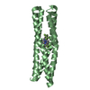
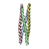
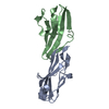
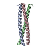
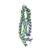
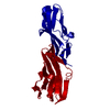
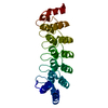
CRYSTAL STRUCTURE OF THE OLIGOMERIZATION DOMAIN FROM ROTAVIRUS NSP4
Components
NON-STRUCTURAL GLYCOPROTEIN NSP4
Keywords
METAL BINDING PROTEIN / NS28 / parallel coiled-coil / homo-tetramer / metal binding site / strontium / cation
Function / homology
Function and homology information
host caveola / host cell rough endoplasmic reticulum membrane / viral process / toxin activity / channel activity / monoatomic ion transmembrane transport / symbiont-mediated activation of host autophagy / extracellular region / membrane / metal ion binding Similarity search - Function
Rotavirus non-structural protein 4 / Rotavirus non structural protein / Single alpha-helices involved in coiled-coils or other helix-helix interfaces - #430 / Single alpha-helices involved in coiled-coils or other helix-helix interfaces / Up-down Bundle / Mainly Alpha Similarity search - Domain/homology
Resolution: 1.86→1.94 Å / Redundancy: 3 % / Rmerge(I) obs: 0.117 / % possible all: 100
Reflection
*PLUS
Num. measured all: 138038
-
Processing
Software
Name
Classification
DENZO
datareduction
SCALEPACK
datascaling
CNS
refinement
CNS
phasing
Refinement
Method to determine structure: MAD / Resolution: 1.86→19.34 Å / Rfactor Rfree error: 0.007 / Data cutoff high absF: 711648.5 / Data cutoff low absF: 0 / Isotropic thermal model: RESTRAINED / Cross valid method: THROUGHOUT / σ(F): 0 / σ(I): 0 / Stereochemistry target values: Engh & Huber Details: Four water molecules have been modelled above the metal and hydrating water molecules. It is also possible that the four water molecules (two per ASU) are instead a small molecule lying ...Details: Four water molecules have been modelled above the metal and hydrating water molecules. It is also possible that the four water molecules (two per ASU) are instead a small molecule lying across the two-fold crystallographic axis. There are five residues whose side chains have alternative rotomer conformations and have been modelled as such: A98, A106, A112, B99, and B114.
In the structure databanks used in Yorodumi, some data are registered as the other names, "COVID-19 virus" and "2019-nCoV". Here are the details of the virus and the list of structure data.
Jan 31, 2019. EMDB accession codes are about to change! (news from PDBe EMDB page)
EMDB accession codes are about to change! (news from PDBe EMDB page)
The allocation of 4 digits for EMDB accession codes will soon come to an end. Whilst these codes will remain in use, new EMDB accession codes will include an additional digit and will expand incrementally as the available range of codes is exhausted. The current 4-digit format prefixed with “EMD-” (i.e. EMD-XXXX) will advance to a 5-digit format (i.e. EMD-XXXXX), and so on. It is currently estimated that the 4-digit codes will be depleted around Spring 2019, at which point the 5-digit format will come into force.
The EM Navigator/Yorodumi systems omit the EMD- prefix.
Related info.:Q: What is EMD? / ID/Accession-code notation in Yorodumi/EM Navigator
Yorodumi is a browser for structure data from EMDB, PDB, SASBDB, etc.
This page is also the successor to EM Navigator detail page, and also detail information page/front-end page for Omokage search.
The word "yorodu" (or yorozu) is an old Japanese word meaning "ten thousand". "mi" (miru) is to see.
Related info.:EMDB / PDB / SASBDB / Comparison of 3 databanks / Yorodumi Search / Aug 31, 2016. New EM Navigator & Yorodumi / Yorodumi Papers / Jmol/JSmol / Function and homology information / Changes in new EM Navigator and Yorodumi
 Movie
Movie Controller

 Yorodumi
 Open data
 Basic information
 Components
 Keywords
 Function and homology information
 X-RAY DIFFRACTION /
  Authors
 Citation
 Structure visualization
 Downloads & links
 Other downloads
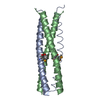


 PDBj
PDBj
 Assembly


 Mass: 87.620 Da / Num. of mol.: 1 / Source method: obtained synthetically / Formula: Sr
 Mass: 18.015 Da / Num. of mol.: 88 / Source method: isolated from a natural source / Formula: H2O
 Sample preparation
 / Beamline: X12C / Wavelength: 0.9785,0.9788,0.95
 Processing