proteasome regulatory particle assembly / positive regulation of cyclin-dependent protein serine/threonine kinase activity / sperm glycocalyx / Proteasome assembly / perinuclear theca / negative regulation of release of cytochrome c from mitochondria / negative regulation of DNA damage response, signal transduction by p53 class mediator / negative regulation of MAPK cascade / ciliary tip / proteasome complex ...proteasome regulatory particle assembly / positive regulation of cyclin-dependent protein serine/threonine kinase activity / sperm glycocalyx / Proteasome assembly / perinuclear theca / negative regulation of release of cytochrome c from mitochondria / negative regulation of DNA damage response, signal transduction by p53 class mediator / negative regulation of MAPK cascade / ciliary tip / proteasome complex / positive regulation of protein ubiquitination / positive regulation of proteasomal ubiquitin-dependent protein catabolic process / sperm principal piece / microtubule cytoskeleton / positive regulation of cell growth / sperm midpiece / cytoskeleton / RNA polymerase II-specific DNA-binding transcription factor binding / cilium / apoptotic process / regulation of transcription by RNA polymerase II / negative regulation of apoptotic process / negative regulation of transcription by RNA polymerase II / nucleus / cytoplasm / cytosol Similarity search - Function
Mass: 24459.855 Da / Num. of mol.: 1 Source method: isolated from a genetically manipulated source Source: (gene. exp.) HOMO SAPIENS (human) / Production host: ESCHERICHIA COLI (E. coli) / Strain (production host): ROSETTA BLUE / References: UniProt: O75832
Mass: 18.015 Da / Num. of mol.: 115 / Source method: isolated from a natural source / Formula: H2O
Compound details
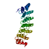
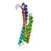
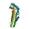
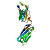
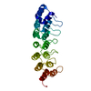
FUNCTIONS AS A REGULATORY SUBUNIT OF THE 26S PROTEASOME INVOLVED IN THE ATP-DEPENDENT DEGRADATION ...FUNCTIONS AS A REGULATORY SUBUNIT OF THE 26S PROTEASOME INVOLVED IN THE ATP-DEPENDENT DEGRADATION OF UBIQUITINATED PROTEINS. A PA700 COMPLEX COMPONENT WITH 5 ANKYRIN REPEATS
-
Experimental details
-
Experiment
Experiment
Method: X-RAY DIFFRACTION / Number of used crystals: 1
-
Sample preparation
Crystal
Density Matthews: 1.8 Å3/Da / Density % sol: 32.6 %
Crystal grow
pH: 6.4 / Details: 24% PEG 5000 MONOMETHYL ETHER, 0.1 M MES PH 6.4
Resolution: 2→32.44 Å / Cor.coef. Fo:Fc: 0.949 / Cor.coef. Fo:Fc free: 0.908 / SU B: 4.934 / SU ML: 0.141 / TLS residual ADP flag: LIKELY RESIDUAL / Cross valid method: THROUGHOUT / ESU R: 0.241 / ESU R Free: 0.201 / Stereochemistry target values: MAXIMUM LIKELIHOOD Details: HYDROGENS HAVE BEEN ADDED IN THE RIDING POSITIONS. THIS ENTRY CONTAINS RESIDUES WITH ZERO OCCUPANCY. THESE RESIDUES ARE A24, A30, A38, A87, A171, A189, A215, A221, A222, A224 AND A226
Rfactor
Num. reflection
% reflection
Selection details
Rfree
0.253
612
4.9 %
RANDOM
Rwork
0.191
-
-
-
obs
0.194
11860
97.2 %
-
Solvent computation
Ion probe radii: 0.8 Å / Shrinkage radii: 0.8 Å / VDW probe radii: 1.4 Å / Solvent model: BABINET MODEL PLUS MASK
 Movie
Movie Controller


 Open data
 Basic information
 Components
 Keywords
 Function and homology information
 HOMO SAPIENS (human)
 X-RAY DIFFRACTION /
  Authors
 Citation
 Structure visualization
 Downloads & links
 Other downloads
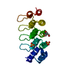

 PDBj
PDBj

 Assembly
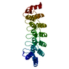

 Mass: 18.015 Da / Num. of mol.: 115 / Source method: isolated from a natural source / Formula: H2O
 Sample preparation
 / Beamline: ID29 / Wavelength: 0.9756
  Processing