 Movie
Movie Controller


+ Open data

- Basic information
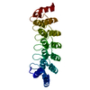
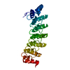
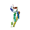
| Entry | Database: PDB / ID: 1s2x | ||||||
|---|---|---|---|---|---|---|---|
| Title | Crystal structure of Cag-Z from Helicobacter pylori | ||||||
 Components Components | Cag-Z | ||||||
 Keywords Keywords | UNKNOWN FUNCTION / Cag-Z / Helicobacter pylori / cag pathogenicity island / type IV secretion system | ||||||
| Function / homology | CAG pathogenicity island protein, CagZ / Cag pathogenicity island protein, CagZ / Cag-Z superfamily / CagZ / Delta-Endotoxin; domain 1 / Up-down Bundle / Mainly Alpha / ISOPROPYL ALCOHOL / Cag-Z Function and homology information Function and homology information | ||||||
| Biological species |   Helicobacter pylori (bacteria) Helicobacter pylori (bacteria) | ||||||
| Method |  X-RAY DIFFRACTION / SYNCHROTRON / MAD / Resolution: 1.9 Å X-RAY DIFFRACTION / SYNCHROTRON / MAD / Resolution: 1.9 Å | ||||||
 Authors Authors | Cendron, L. / Seydel, A. / Angelini, A. / Battistutta, R. / Zanotti, G. | ||||||
 Citation Citation | Journal: J.Mol.Biol. / Year: 2004 Title: Crystal structure of CagZ, a protein from the Helicobacter pylori pathogenicity island that encodes for a type IV secretion system Authors: Cendron, L. / Seydel, A. / Angelini, A. / Battistutta, R. / Zanotti, G. | ||||||
| History |
|
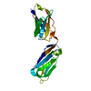
- Structure visualization
| Structure viewer | Molecule: MolmilJmol/JSmol |
|---|
- Downloads & links
-Download
| PDBx/mmCIF format | 1s2x.cif.gz | 52 KB | Display | PDBx/mmCIF format |
|---|---|---|---|---|
| PDB format | pdb1s2x.ent.gz | 36.5 KB | Display | PDB format |
| PDBx/mmJSON format | 1s2x.json.gz | Tree view | PDBx/mmJSON format | |
| Others |  Other downloads Other downloads |
-Validation report
| Arichive directory | https://data.pdbj.org/pub/pdb/validation_reports/s2/1s2xftp://data.pdbj.org/pub/pdb/validation_reports/s2/1s2x | HTTPS FTP |
|---|
-Related structure data
| Similar structure data |
|---|


-Links
 PDBj
PDBj

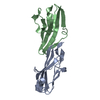
- Assembly
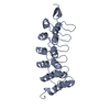
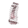
| Deposited unit | 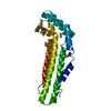
| ||||||||
|---|---|---|---|---|---|---|---|---|---|
| 1 |
| ||||||||
| Unit cell |
|
-Components
| #1: Protein | Mass: 23938.371 Da / Num. of mol.: 1 / Mutation: M1D Source method: isolated from a genetically manipulated source Source: (gene. exp.) Helicobacter pylori (bacteria) / Gene: cagZ / Plasmid: pGEX-4T-3 / Species (production host): Escherichia coli / Production host: |
|---|---|
| #2: Chemical | ChemComp-IPA /   Mass: 60.095 Da / Num. of mol.: 1 / Source method: obtained synthetically / Formula: C3H8O / Comment: alkaloid*YM Mass: 60.095 Da / Num. of mol.: 1 / Source method: obtained synthetically / Formula: C3H8O / Comment: alkaloid*YM |
| #3: Water | ChemComp-HOH /  Mass: 18.015 Da / Num. of mol.: 91 / Source method: isolated from a natural source / Formula: H2O Mass: 18.015 Da / Num. of mol.: 91 / Source method: isolated from a natural source / Formula: H2O |
| Has protein modification | Y |
-Experimental details
-Experiment
| Experiment | Method: X-RAY DIFFRACTION / Number of used crystals: 1 |
|---|
- Sample preparation
| Crystal | Density Matthews: 2.74 Å3/Da / Density % sol: 54.7 % |
|---|---|
| Crystal grow | Temperature: 298 K / Method: vapor diffusion, sitting drop / pH: 7.5 Details: PEG 4000, isopropanol, HEPES, pH 7.5, VAPOR DIFFUSION, SITTING DROP, temperature 298K |
-Data collection
| Diffraction | Mean temperature: 100 K |
|---|---|
| Diffraction source | Source: SYNCHROTRON / Site: ESRF  / Beamline: ID29 / Wavelength: 0.9202 Å / Beamline: ID29 / Wavelength: 0.9202 Å |
| Detector | Type: ADSC QUANTUM 4 / Detector: CCD / Date: Sep 10, 2003 |
| Radiation | Protocol: MAD / Monochromatic (M) / Laue (L): M / Scattering type: x-ray |
| Radiation wavelength | Wavelength: 0.9202 Å / Relative weight: 1 |
| Reflection | Resolution: 1.9→45 Å / Num. all: 18377 / Num. obs: 18377 / % possible obs: 98.6 % / Observed criterion σ(F): 0 / Observed criterion σ(I): 0 / Redundancy: 6.2 % / Rmerge(I) obs: 0.054 / Net I/σ(I): 5 |
| Reflection shell | Resolution: 1.9→2 Å / Redundancy: 4.1 % / Rmerge(I) obs: 0.246 / Mean I/σ(I) obs: 1.7 / Num. unique all: 2406 / % possible all: 92.5 |
- Processing
| Software |
| |||||||||||||||||||||||||||||||||||
|---|---|---|---|---|---|---|---|---|---|---|---|---|---|---|---|---|---|---|---|---|---|---|---|---|---|---|---|---|---|---|---|---|---|---|---|---|
| Refinement | Method to determine structure: MAD / Resolution: 1.9→8 Å / Num. parameters: 6267 / Num. restraintsaints: 5971 Isotropic thermal model: Isotropic, Anisotropic only for 4 Se atoms Cross valid method: FREE R / σ(F): 2 / σ(I): 4 / Stereochemistry target values: ENGH AND HUBER Details: ANISOTROPIC SCALING APPLIED BY THE METHOD OF PARKIN, MOEZZI & HOPE, J.APPL.CRYST.28(1995)53-56
| |||||||||||||||||||||||||||||||||||
| Refine analyze | Num. disordered residues: 0 / Occupancy sum hydrogen: 0 / Occupancy sum non hydrogen: 1556 | |||||||||||||||||||||||||||||||||||
| Refinement step | Cycle: LAST / Resolution: 1.9→8 Å
| |||||||||||||||||||||||||||||||||||
| Refine LS restraints |
| |||||||||||||||||||||||||||||||||||
| LS refinement shell |
|
