 Movie
Movie Controller

[English] 日本語
 Yorodumi
- PDB-2wk4: Dimeric structure of D347G D348G mutant of the sapporovirus RNA d... -
+ Open data

- Basic information
| Entry | Database: PDB / ID: 2wk4 | ||||||
|---|---|---|---|---|---|---|---|
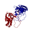
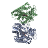
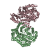
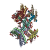
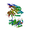
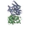
| Title | Dimeric structure of D347G D348G mutant of the sapporovirus RNA dependent RNA polymerase | ||||||
 Components Components | PROTEASE-POLYMERASE P70 | ||||||
 Keywords Keywords | HYDROLASE / COVALENT PROTEIN-RNA LINKAGE / NUCLEOTIDYLTRANSFERASE / RNA REPLICATION / PHOSPHORYLATION / RNA ELONGATION / THIOL PROTEASE / CAPSID PROTEIN / ATP-BINDING / HELICASE / PROTEASE | ||||||
| Function / homology |  Function and homology information Function and homology informationcalicivirin / ribonucleoside triphosphate phosphatase activity / viral capsid / nucleoside-triphosphate phosphatase / host cell cytoplasm / DNA replication / RNA helicase activity / RNA-directed RNA polymerase / cysteine-type endopeptidase activity / viral RNA genome replication ...calicivirin / ribonucleoside triphosphate phosphatase activity / viral capsid / nucleoside-triphosphate phosphatase / host cell cytoplasm / DNA replication / RNA helicase activity / RNA-directed RNA polymerase / cysteine-type endopeptidase activity / viral RNA genome replication / RNA-directed RNA polymerase activity / DNA-templated transcription / proteolysis / RNA binding / ATP binding Similarity search - Function | ||||||
| Biological species |  SAPPORO VIRUS SAPPORO VIRUS | ||||||
| Method |  X-RAY DIFFRACTION / SYNCHROTRON / MOLECULAR REPLACEMENT / Resolution: 2.98 Å X-RAY DIFFRACTION / SYNCHROTRON / MOLECULAR REPLACEMENT / Resolution: 2.98 Å | ||||||
 Authors Authors | Fullerton, S.W.B. / Robel, I. / Schuldt, L. / Gebhardt, J. / Tucker, P.A. / Rohayem, J. | ||||||
 Citation Citation | Journal: To be Published Title: Dimeric Structure of D347G D348G Mutant of the Sapporovirus Sapporovirus RNA Dependent RNA Polymerase Authors: Fullerton, S.W.B. / Robel, I. / Schuldt, L. / Gebhardt, J. / Tucker, P.A. / Rohayem, J. | ||||||
| History |
|
- Structure visualization
| Structure viewer | Molecule: MolmilJmol/JSmol |
|---|
- Downloads & links
-Download
| PDBx/mmCIF format | 2wk4.cif.gz | 201.9 KB | Display | PDBx/mmCIF format |
|---|---|---|---|---|
| PDB format | pdb2wk4.ent.gz | 162.3 KB | Display | PDB format |
| PDBx/mmJSON format | 2wk4.json.gz | Tree view | PDBx/mmJSON format | |
| Others |  Other downloads Other downloads |
-Validation report
| Arichive directory | https://data.pdbj.org/pub/pdb/validation_reports/wk/2wk4ftp://data.pdbj.org/pub/pdb/validation_reports/wk/2wk4 | HTTPS FTP |
|---|
-Related structure data
| Related structure data | 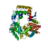 2ckwS S: Starting model for refinement |
|---|---|
| Similar structure data |


-Links
 PDBj
PDBj

- Assembly
| Deposited unit | 
| ||||||||||||||||||
|---|---|---|---|---|---|---|---|---|---|---|---|---|---|---|---|---|---|---|---|
| 1 |
| ||||||||||||||||||
| Unit cell |
| ||||||||||||||||||
| Noncrystallographic symmetry (NCS) | NCS domain:
NCS domain segments: Component-ID: 1 / Ens-ID: 1 / Beg auth comp-ID: LYS / Beg label comp-ID: LYS / End auth comp-ID: GLY / End label comp-ID: GLY / Refine code: 6 / Auth seq-ID: 12 - 502 / Label seq-ID: 12 - 502
NCS oper: (Code: given Matrix: (-0.9097, -0.3897, 0.1432), Vector: |
-Components
| #1: Protein | Mass: 56369.633 Da / Num. of mol.: 2 / Fragment: RESIDUES 1135-1649 / Mutation: YES Source method: isolated from a genetically manipulated source Source: (gene. exp.) SAPPORO VIRUS / Production host:  #2: Chemical |   Mass: 92.094 Da / Num. of mol.: 3 / Source method: obtained synthetically / Formula: C3H8O3 Mass: 92.094 Da / Num. of mol.: 3 / Source method: obtained synthetically / Formula: C3H8O3#3: Water | ChemComp-HOH / |  Mass: 18.015 Da / Num. of mol.: 209 / Source method: isolated from a natural source / Formula: H2O Mass: 18.015 Da / Num. of mol.: 209 / Source method: isolated from a natural source / Formula: H2OCompound details | ENGINEERED RESIDUE IN CHAIN A, ASP 1481 TO GLY ENGINEERED RESIDUE IN CHAIN A, ASP 1482 TO GLY ...ENGINEERED | |
|---|
-Experimental details
-Experiment
| Experiment | Method: X-RAY DIFFRACTION |
|---|
- Sample preparation
| Crystal | Density Matthews: 2.99 Å3/Da / Density % sol: 58.49 % / Description: NONE |
|---|
-Data collection
| Diffraction | Mean temperature: 100 K |
|---|---|
| Diffraction source | Source: SYNCHROTRON / Site: EMBL/DESY, HAMBURG  / Beamline: X11 / Wavelength: 0.81 / Beamline: X11 / Wavelength: 0.81 |
| Detector | Type: MAR555 FLAT PANEL / Detector: IMAGE PLATE / Details: MIRRORS |
| Radiation | Protocol: SINGLE WAVELENGTH / Monochromatic (M) / Laue (L): M / Scattering type: x-ray |
| Radiation wavelength | Wavelength: 0.81 Å / Relative weight: 1 |
| Reflection | Resolution: 2.9→50 Å / Num. obs: 29506 / % possible obs: 96 % / Observed criterion σ(I): -3 / Redundancy: 7.4 % / Biso Wilson estimate: 61.1 Å2 / Rmerge(I) obs: 0.21 / Net I/σ(I): 10.8 |
| Reflection shell | Resolution: 2.9→3.06 Å / Redundancy: 6.3 % / Rmerge(I) obs: 0.99 / Mean I/σ(I) obs: 2 / % possible all: 72.8 |
- Processing
| Software |
| ||||||||||||||||||||||||||||||||||||||||||||||||||||||||||||||||||||||||||||||||||||||||||||||||||||||||||||||||||||||||||||||||||||||||||||||||||||||||||||||||||||||||||||||||||||||
|---|---|---|---|---|---|---|---|---|---|---|---|---|---|---|---|---|---|---|---|---|---|---|---|---|---|---|---|---|---|---|---|---|---|---|---|---|---|---|---|---|---|---|---|---|---|---|---|---|---|---|---|---|---|---|---|---|---|---|---|---|---|---|---|---|---|---|---|---|---|---|---|---|---|---|---|---|---|---|---|---|---|---|---|---|---|---|---|---|---|---|---|---|---|---|---|---|---|---|---|---|---|---|---|---|---|---|---|---|---|---|---|---|---|---|---|---|---|---|---|---|---|---|---|---|---|---|---|---|---|---|---|---|---|---|---|---|---|---|---|---|---|---|---|---|---|---|---|---|---|---|---|---|---|---|---|---|---|---|---|---|---|---|---|---|---|---|---|---|---|---|---|---|---|---|---|---|---|---|---|---|---|---|---|
| Refinement | Method to determine structure: MOLECULAR REPLACEMENT Starting model: PDB ENTRY 2CKW Resolution: 2.98→26.01 Å / Cor.coef. Fo:Fc: 0.927 / Cor.coef. Fo:Fc free: 0.837 / SU B: 18.818 / SU ML: 0.353 / Cross valid method: THROUGHOUT / ESU R Free: 0.484 / Stereochemistry target values: MAXIMUM LIKELIHOOD Details: HYDROGENS HAVE BEEN ADDED IN THE RIDING POSITIONS. U VALUES REFINED INDIVIDUALLY
| ||||||||||||||||||||||||||||||||||||||||||||||||||||||||||||||||||||||||||||||||||||||||||||||||||||||||||||||||||||||||||||||||||||||||||||||||||||||||||||||||||||||||||||||||||||||
| Solvent computation | Ion probe radii: 0.8 Å / Shrinkage radii: 0.8 Å / VDW probe radii: 1.4 Å / Solvent model: MASK | ||||||||||||||||||||||||||||||||||||||||||||||||||||||||||||||||||||||||||||||||||||||||||||||||||||||||||||||||||||||||||||||||||||||||||||||||||||||||||||||||||||||||||||||||||||||
| Displacement parameters | Biso mean: 38.478 Å2
| ||||||||||||||||||||||||||||||||||||||||||||||||||||||||||||||||||||||||||||||||||||||||||||||||||||||||||||||||||||||||||||||||||||||||||||||||||||||||||||||||||||||||||||||||||||||
| Refinement step | Cycle: LAST / Resolution: 2.98→26.01 Å
| ||||||||||||||||||||||||||||||||||||||||||||||||||||||||||||||||||||||||||||||||||||||||||||||||||||||||||||||||||||||||||||||||||||||||||||||||||||||||||||||||||||||||||||||||||||||
| Refine LS restraints |
|