 Movie
Movie Controller


+ Open data

- Basic information
| Entry | Database: PDB / ID: 1og2 | ||||||
|---|---|---|---|---|---|---|---|
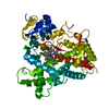
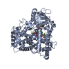
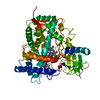
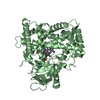
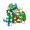
| Title | Structure of human cytochrome P450 CYP2C9 | ||||||
 Components Components | CYTOCHROME P450 2C9 | ||||||
 Keywords Keywords | ELECTRON TRANSPORT / DRUG METABOLISM / OXIDOREDUCTASE / HEME / MONOOXYGENASE | ||||||
| Function / homology |  Function and homology information Function and homology informationarachidonate 14,15-epoxygenase activity / arachidonate 11,12-epoxygenase activity / (S)-limonene 6-monooxygenase / (S)-limonene 7-monooxygenase / (R)-limonene 6-monooxygenase / (S)-limonene 6-monooxygenase activity / (S)-limonene 7-monooxygenase activity / (R)-limonene 6-monooxygenase activity / Synthesis of (16-20)-hydroxyeicosatetraenoic acids (HETE) / omega-hydroxylase P450 pathway ...arachidonate 14,15-epoxygenase activity / arachidonate 11,12-epoxygenase activity / (S)-limonene 6-monooxygenase / (S)-limonene 7-monooxygenase / (R)-limonene 6-monooxygenase / (S)-limonene 6-monooxygenase activity / (S)-limonene 7-monooxygenase activity / (R)-limonene 6-monooxygenase activity / Synthesis of (16-20)-hydroxyeicosatetraenoic acids (HETE) / omega-hydroxylase P450 pathway / organofluorine metabolic process / CYP2E1 reactions / arachidonate epoxygenase activity / epoxygenase P450 pathway / monocarboxylic acid metabolic process / icosanoid biosynthetic process / urea metabolic process / caffeine oxidase activity / Biosynthesis of maresin-like SPMs / monoterpenoid metabolic process / Synthesis of epoxy (EET) and dihydroxyeicosatrienoic acids (DHET) / estrogen 2-hydroxylase activity / oxidative demethylation / steroid hydroxylase activity / Xenobiotics / oxidoreductase activity, acting on paired donors, with incorporation or reduction of molecular oxygen, reduced flavin or flavoprotein as one donor, and incorporation of one atom of oxygen / estrogen metabolic process / unspecific monooxygenase / long-chain fatty acid biosynthetic process / Aspirin ADME / steroid metabolic process / intracellular membrane-bounded organelle / xenobiotic catabolic process / cholesterol metabolic process / xenobiotic metabolic process / monooxygenase activity / oxidoreductase activity / iron ion binding / heme binding / endoplasmic reticulum membrane / plasma membrane / cytoplasm Similarity search - Function | ||||||
| Biological species |  HOMO SAPIENS (human) HOMO SAPIENS (human) | ||||||
| Method |  X-RAY DIFFRACTION / SYNCHROTRON / MOLECULAR REPLACEMENT / Resolution: 2.6 Å X-RAY DIFFRACTION / SYNCHROTRON / MOLECULAR REPLACEMENT / Resolution: 2.6 Å | ||||||
 Authors Authors | Williams, P.A. / Cosme, J. / Ward, A. / Angove, H.C. / Matak Vinkovic, D. / Jhoti, H. | ||||||
 Citation Citation | Journal: Nature / Year: 2003 Title: Crystal Structure of Human Cytochrome P450 2C9 with Bound Warfarin Authors: Williams, P.A. / Cosme, J. / Ward, A. / Angove, H.C. / Matak Vinkovic, D. / Jhoti, H. | ||||||
| History |
|
- Structure visualization
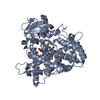
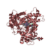
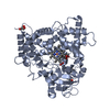
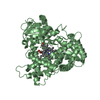
| Structure viewer | Molecule: MolmilJmol/JSmol |
|---|
- Downloads & links
-Download
| PDBx/mmCIF format | 1og2.cif.gz | 198.8 KB | Display | PDBx/mmCIF format |
|---|---|---|---|---|
| PDB format | pdb1og2.ent.gz | 159.2 KB | Display | PDB format |
| PDBx/mmJSON format | 1og2.json.gz | Tree view | PDBx/mmJSON format | |
| Others |  Other downloads Other downloads |
-Validation report
| Arichive directory | https://data.pdbj.org/pub/pdb/validation_reports/og/1og2ftp://data.pdbj.org/pub/pdb/validation_reports/og/1og2 | HTTPS FTP |
|---|
-Related structure data
| Related structure data |  1og5C 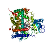 1dt6S S: Starting model for refinement C: citing same article ( |
|---|---|
| Similar structure data |

-Links
 PDBj
PDBj


- Assembly
| Deposited unit | 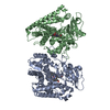
| ||||||||
|---|---|---|---|---|---|---|---|---|---|
| 1 | 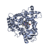
| ||||||||
| 2 | 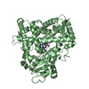
| ||||||||
| Unit cell |
| ||||||||
| Components on special symmetry positions |
| ||||||||
| Noncrystallographic symmetry (NCS) | NCS oper: (Code: given Matrix: (-0.8191, 0.5736, 0.0033), Vector: |
-Components
| #1: Protein | Mass: 54168.590 Da / Num. of mol.: 2 / Fragment: SOLUBLE DOMAIN, RESIDUES 30-490 / Mutation: YES Source method: isolated from a genetically manipulated source Source: (gene. exp.) HOMO SAPIENS (human) / Production host:  References: UniProt: P11712, EC: 1.14.13.80, EC: 1.14.13.48, EC: 1.14.13.49 #2: Chemical |   Mass: 618.503 Da / Num. of mol.: 2 / Source method: obtained synthetically / Formula: C34H34FeN4O4 Mass: 618.503 Da / Num. of mol.: 2 / Source method: obtained synthetically / Formula: C34H34FeN4O4#3: Water | ChemComp-HOH / |  Mass: 18.015 Da / Num. of mol.: 147 / Source method: isolated from a natural source / Formula: H2O Mass: 18.015 Da / Num. of mol.: 147 / Source method: isolated from a natural source / Formula: H2OCompound details | CYTOCHROMES P450 ARE HEME-THIOLATE MONOOXYGENASES THAT IN THE LIVER MICROSOMES FORM PART OF THE ...CYTOCHROME | Sequence details | RESIDUES 1-29 OF THE FULL LENGTH PROTEIN WERE DELETED (THE PROPOSED TRANSMEMBRANE DOMAIN) AND A ...RESIDUES 1-29 OF THE FULL LENGTH PROTEIN WERE DELETED (THE PROPOSED TRANSMEMBR | |
|---|
-Experimental details
-Experiment
| Experiment | Method: X-RAY DIFFRACTION / Number of used crystals: 1 |
|---|
- Sample preparation
| Crystal | Density Matthews: 4.02 Å3/Da / Density % sol: 70 % | |||||||||||||||||||||||||||||||||||||||||||||||||||||||||||||||||||||||||||||
|---|---|---|---|---|---|---|---|---|---|---|---|---|---|---|---|---|---|---|---|---|---|---|---|---|---|---|---|---|---|---|---|---|---|---|---|---|---|---|---|---|---|---|---|---|---|---|---|---|---|---|---|---|---|---|---|---|---|---|---|---|---|---|---|---|---|---|---|---|---|---|---|---|---|---|---|---|---|---|
| Crystal grow | pH: 8.4 / Details: pH 8.40 | |||||||||||||||||||||||||||||||||||||||||||||||||||||||||||||||||||||||||||||
| Crystal grow | *PLUS Temperature: 25 ℃ / pH: 7.4 / Method: vapor diffusion, hanging drop | |||||||||||||||||||||||||||||||||||||||||||||||||||||||||||||||||||||||||||||
| Components of the solutions | *PLUS
|
-Data collection
| Diffraction | Mean temperature: 100 K |
|---|---|
| Diffraction source | Source: SYNCHROTRON / Site: ESRF  / Beamline: ID14-2 / Wavelength: 0.9 / Beamline: ID14-2 / Wavelength: 0.9 |
| Detector | Type: ADSC CCD / Detector: CCD |
| Radiation | Protocol: SINGLE WAVELENGTH / Monochromatic (M) / Laue (L): M / Scattering type: x-ray |
| Radiation wavelength | Wavelength: 0.9 Å / Relative weight: 1 |
| Reflection | Resolution: 2.6→50 Å / Num. obs: 51912 / % possible obs: 96.5 % / Redundancy: 2.6 % / Rmerge(I) obs: 0.087 / Net I/σ(I): 6.8 |
| Reflection shell | Resolution: 2.6→2.74 Å / Redundancy: 2 % / Rmerge(I) obs: 0.57 / Mean I/σ(I) obs: 1.21 / % possible all: 96.5 |
| Reflection | *PLUS Highest resolution: 2.6 Å / Lowest resolution: 50 Å / Redundancy: 2.6 % / Num. measured all: 340743 / Rmerge(I) obs: 0.087 |
| Reflection shell | *PLUS Highest resolution: 2.6 Å / % possible obs: 96.5 % / Redundancy: 2 % / Rmerge(I) obs: 0.57 / Mean I/σ(I) obs: 1.21 |
- Processing
| Software |
| ||||||||||||||||||||||||||||||||||||||||||||||||||||||||||||||||||||||||||||||||||||||||||||||||||||||||||||||||||||||||||||||||||||||||||||||||||||||||||||||||||||||||||||||||||||||
|---|---|---|---|---|---|---|---|---|---|---|---|---|---|---|---|---|---|---|---|---|---|---|---|---|---|---|---|---|---|---|---|---|---|---|---|---|---|---|---|---|---|---|---|---|---|---|---|---|---|---|---|---|---|---|---|---|---|---|---|---|---|---|---|---|---|---|---|---|---|---|---|---|---|---|---|---|---|---|---|---|---|---|---|---|---|---|---|---|---|---|---|---|---|---|---|---|---|---|---|---|---|---|---|---|---|---|---|---|---|---|---|---|---|---|---|---|---|---|---|---|---|---|---|---|---|---|---|---|---|---|---|---|---|---|---|---|---|---|---|---|---|---|---|---|---|---|---|---|---|---|---|---|---|---|---|---|---|---|---|---|---|---|---|---|---|---|---|---|---|---|---|---|---|---|---|---|---|---|---|---|---|---|---|
| Refinement | Method to determine structure: MOLECULAR REPLACEMENT Starting model: PDB ENTRY 1DT6 Resolution: 2.6→55.9 Å / Cor.coef. Fo:Fc: 0.936 / Cor.coef. Fo:Fc free: 0.909 / SU B: 10.96 / SU ML: 0.217 / Cross valid method: THROUGHOUT / ESU R: 0.354 / ESU R Free: 0.273 / Stereochemistry target values: MAXIMUM LIKELIHOOD / Details: HYDROGENS HAVE BEEN ADDED IN THE RIDING POSITIONS
| ||||||||||||||||||||||||||||||||||||||||||||||||||||||||||||||||||||||||||||||||||||||||||||||||||||||||||||||||||||||||||||||||||||||||||||||||||||||||||||||||||||||||||||||||||||||
| Solvent computation | Ion probe radii: 0.8 Å / Shrinkage radii: 0.8 Å / VDW probe radii: 1.4 Å / Solvent model: BABINET MODEL WITH MASK | ||||||||||||||||||||||||||||||||||||||||||||||||||||||||||||||||||||||||||||||||||||||||||||||||||||||||||||||||||||||||||||||||||||||||||||||||||||||||||||||||||||||||||||||||||||||
| Displacement parameters | Biso mean: 47.75 Å2
| ||||||||||||||||||||||||||||||||||||||||||||||||||||||||||||||||||||||||||||||||||||||||||||||||||||||||||||||||||||||||||||||||||||||||||||||||||||||||||||||||||||||||||||||||||||||
| Refinement step | Cycle: LAST / Resolution: 2.6→55.9 Å
| ||||||||||||||||||||||||||||||||||||||||||||||||||||||||||||||||||||||||||||||||||||||||||||||||||||||||||||||||||||||||||||||||||||||||||||||||||||||||||||||||||||||||||||||||||||||
| Refine LS restraints |
|