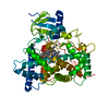
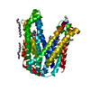
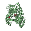
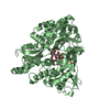
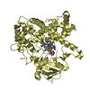
[English] 日本語
Yorodumi
- PDB-5a5i: Cytochrome 2C9 P450 inhibitor complex -

+
Open data


ID or keywords:

Loading...

-
Basic information

Entry
Database: PDB / ID: 5a5i
TitleCytochrome 2C9 P450 inhibitor complex
ComponentsCYTOCHROME P450 2C9
KeywordsOXIDOREDUCTASE
Function / homology
Function and homology information


arachidonic acid 14,15-epoxygenase activity / arachidonic acid 11,12-epoxygenase activity / amide metabolic process / (S)-limonene 6-monooxygenase / (S)-limonene 7-monooxygenase / (R)-limonene 6-monooxygenase / (S)-limonene 6-monooxygenase activity / (S)-limonene 7-monooxygenase activity / (R)-limonene 6-monooxygenase activity / monocarboxylic acid metabolic process ...arachidonic acid 14,15-epoxygenase activity / arachidonic acid 11,12-epoxygenase activity / amide metabolic process / (S)-limonene 6-monooxygenase / (S)-limonene 7-monooxygenase / (R)-limonene 6-monooxygenase / (S)-limonene 6-monooxygenase activity / (S)-limonene 7-monooxygenase activity / (R)-limonene 6-monooxygenase activity / monocarboxylic acid metabolic process / urea metabolic process / Synthesis of (16-20)-hydroxyeicosatetraenoic acids (HETE) / omega-hydroxylase P450 pathway / CYP2E1 reactions / arachidonic acid epoxygenase activity / icosanoid biosynthetic process / epoxygenase P450 pathway / caffeine oxidase activity / Biosynthesis of maresin-like SPMs / Synthesis of epoxy (EET) and dihydroxyeicosatrienoic acids (DHET) / monoterpenoid metabolic process / oxidative demethylation / Xenobiotics / oxidoreductase activity, acting on paired donors, with incorporation or reduction of molecular oxygen, reduced flavin or flavoprotein as one donor, and incorporation of one atom of oxygen / long-chain fatty acid biosynthetic process / estrogen metabolic process / unspecific monooxygenase / aromatase activity / Aspirin ADME / steroid metabolic process / steroid hydroxylase activity / xenobiotic catabolic process / monooxygenase activity / cholesterol metabolic process / xenobiotic metabolic process / oxidoreductase activity / iron ion binding / intracellular membrane-bounded organelle / heme binding / endoplasmic reticulum membrane / plasma membrane / cytoplasm
Similarity search - Function
Cytochrome P450, E-class, group I / Cytochrome p450 / Cytochrome P450 / Cytochrome P450, conserved site / Cytochrome P450 cysteine heme-iron ligand signature. / Cytochrome P450 / Cytochrome P450 superfamily / Cytochrome P450 / Orthogonal Bundle / Mainly Alpha
Similarity search - Domain/homology
PROTOPORPHYRIN IX CONTAINING FE / Chem-XI1 / Cytochrome P450 2C9
Similarity search - Component
Biological speciesHOMO SAPIENS (human)
MethodX-RAY DIFFRACTION / SYNCHROTRON / OTHER / Resolution: 2 Å
AuthorsSkerratt, S.E. / de Groot, M.J. / Phillips, C.
CitationJournal: Med.Chem.Comm. / Year: 2016
Title: Discovery of a Novel Binding Pocket for Cyp 2C9 Inhibitors: Crystallography, Pharmacophore Modelling and Inhibitor Sar.
Authors: Skerratt, S.E. / de Groot, M.J. / Phillips, C.
History
DepositionJun 18, 2015Deposition site: PDBE / Processing site: PDBE
Revision 1.0Aug 24, 2016Provider: repository / Type: Initial release
Revision 1.1May 8, 2024Group: Data collection / Database references ...Data collection / Database references / Derived calculations / Other
Category: chem_comp_atom / chem_comp_bond ...chem_comp_atom / chem_comp_bond / citation_author / database_2 / pdbx_database_status / struct_site
Item: _citation_author.name / _database_2.pdbx_DOI ..._citation_author.name / _database_2.pdbx_DOI / _database_2.pdbx_database_accession / _pdbx_database_status.status_code_sf / _struct_site.pdbx_auth_asym_id / _struct_site.pdbx_auth_comp_id / _struct_site.pdbx_auth_seq_id

-
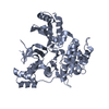
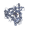
Structure visualization

Structure viewerMolecule:
MolmilJmol/JSmol

Downloads & links

-
Assembly

Deposited unit
A: CYTOCHROME P450 2C9
hetero molecules


Theoretical massNumber of molelcules
Total (without water)55,3223
Polymers54,3231
Non-polymers9992
Water1267
1


  • Idetical with deposited unit
  • defined by software
TypeNameSymmetry operationNumber
identity operation1_555x,y,z1
MethodPISA
Unit cell
Length a, b, c (Å)92.217, 92.217, 170.414
Angle α, β, γ (deg.)90.00, 90.00, 120.00
Int Tables number146
Space group name H-MH3

-
Components

#1: Protein CYTOCHROME P450 2C9 / (R)-LIMONENE 6-MONOOXYGENASE / (S)-LIMONENE 6-MONOOXYGENASE / (S)-LIMONENE 7-MONOOXYGENASE / ...(R)-LIMONENE 6-MONOOXYGENASE / (S)-LIMONENE 6-MONOOXYGENASE / (S)-LIMONENE 7-MONOOXYGENASE / CYPIIC9 / CYTOCHROME P-450MP / CYTOCH ROME P450 MP-4 / CYTOCHROME P450 MP-8 / CYTOCHROME P450 PB-1 / S-MEPH ENYTOIN 4-HYDROXYLASE / CYTOCHROME 2C9


Mass: 54322.887 Da / Num. of mol.: 1 / Fragment: UNP RESIDUES 23-489
Source method: isolated from a genetically manipulated source
Source: (gene. exp.) HOMO SAPIENS (human) / Production host: ESCHERICHIA COLI (E. coli) / References: UniProt: P11712
#2: Chemical ChemComp-XI1 / N-[4-(3-chloranyl-4-cyano-phenoxy)cyclohexyl]-1,1,1-tris(fluoranyl)methanesulfonamide


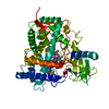
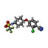
Mass: 382.786 Da / Num. of mol.: 1 / Source method: obtained synthetically / Formula: C14H14ClF3N2O3S
#3: Chemical ChemComp-HEM / PROTOPORPHYRIN IX CONTAINING FE / HEME


Mass: 616.487 Da / Num. of mol.: 1 / Source method: obtained synthetically / Formula: C34H32FeN4O4
#4: Water ChemComp-HOH / water


Mass: 18.015 Da / Num. of mol.: 7 / Source method: isolated from a natural source / Formula: H2O

-
Experimental details

-
Experiment

ExperimentMethod: X-RAY DIFFRACTION

-
Sample preparation

CrystalDensity Matthews: 2.57 Å3/Da / Density % sol: 52.08 % / Description: NONE

-
Data collection

DiffractionMean temperature: 100 K
Diffraction sourceSource: SYNCHROTRON / Site: ESRF / Type: ESRF / Wavelength: 1
RadiationProtocol: SINGLE WAVELENGTH / Monochromatic (M) / Laue (L): M / Scattering type: x-ray
Radiation wavelengthWavelength: 1 Å / Relative weight: 1
ReflectionResolution: 2→25 Å / Num. obs: 27322 / % possible obs: 94.7 % / Observed criterion σ(I): 1 / Redundancy: 2.8 % / Rmerge(I) obs: 0.08

-
Processing

SoftwareName: REFMAC / Version: 5.7.0032 / Classification: refinement
RefinementMethod to determine structure: OTHER
Starting model: NONE

Resolution: 2→24.63 Å / Cor.coef. Fo:Fc: 0.95 / Cor.coef. Fo:Fc free: 0.939 / SU B: 4.992 / SU ML: 0.136 / Cross valid method: THROUGHOUT / ESU R: 0.201 / ESU R Free: 0.171 / Stereochemistry target values: MAXIMUM LIKELIHOOD / Details: HYDROGENS HAVE BEEN ADDED IN THE RIDING POSITIONS
RfactorNum. reflection% reflectionSelection details
Rfree0.24501 1733 5 %RANDOM
Rwork0.2124 ---
obs0.21409 32791 94.57 %-
Solvent computationIon probe radii: 0.8 Å / Shrinkage radii: 0.8 Å / VDW probe radii: 1.2 Å / Solvent model: MASK
Refinement stepCycle: LAST / Resolution: 2→24.63 Å
ProteinNucleic acidLigandSolventTotal
Num. atoms3628 0 67 7 3702
Refine LS restraints
Refine-IDTypeDev idealDev ideal targetNumber
X-RAY DIFFRACTIONr_bond_refined_d0.0170.0193810
X-RAY DIFFRACTIONr_bond_other_d0.0010.023670
X-RAY DIFFRACTIONr_angle_refined_deg1.91.9895166
X-RAY DIFFRACTIONr_angle_other_deg0.89538478
X-RAY DIFFRACTIONr_dihedral_angle_1_deg6.915452
X-RAY DIFFRACTIONr_dihedral_angle_2_deg38.29424.036166
X-RAY DIFFRACTIONr_dihedral_angle_3_deg19.61915672
X-RAY DIFFRACTIONr_dihedral_angle_4_deg20.0861520
X-RAY DIFFRACTIONr_chiral_restr0.1110.2565
X-RAY DIFFRACTIONr_gen_planes_refined0.0090.0214245
X-RAY DIFFRACTIONr_gen_planes_other0.0050.02867
X-RAY DIFFRACTIONr_nbd_refined
X-RAY DIFFRACTIONr_nbd_other
X-RAY DIFFRACTIONr_nbtor_refined
X-RAY DIFFRACTIONr_nbtor_other
X-RAY DIFFRACTIONr_xyhbond_nbd_refined
X-RAY DIFFRACTIONr_xyhbond_nbd_other
X-RAY DIFFRACTIONr_metal_ion_refined
X-RAY DIFFRACTIONr_metal_ion_other
X-RAY DIFFRACTIONr_symmetry_vdw_refined
X-RAY DIFFRACTIONr_symmetry_vdw_other
X-RAY DIFFRACTIONr_symmetry_hbond_refined
X-RAY DIFFRACTIONr_symmetry_hbond_other
X-RAY DIFFRACTIONr_symmetry_metal_ion_refined
X-RAY DIFFRACTIONr_symmetry_metal_ion_other
X-RAY DIFFRACTIONr_mcbond_it3.9964.0571817
X-RAY DIFFRACTIONr_mcbond_other3.9954.0581818
X-RAY DIFFRACTIONr_mcangle_it6.0496.062267
X-RAY DIFFRACTIONr_mcangle_other6.0516.0612267
X-RAY DIFFRACTIONr_scbond_it4.3614.5971993
X-RAY DIFFRACTIONr_scbond_other4.3574.5941987
X-RAY DIFFRACTIONr_scangle_it
X-RAY DIFFRACTIONr_scangle_other6.6356.6822891
X-RAY DIFFRACTIONr_long_range_B_refined9.69732.5774223
X-RAY DIFFRACTIONr_long_range_B_other9.69732.5844224
X-RAY DIFFRACTIONr_rigid_bond_restr
X-RAY DIFFRACTIONr_sphericity_free
X-RAY DIFFRACTIONr_sphericity_bonded
LS refinement shellResolution: 2→2.052 Å / Total num. of bins used: 20
RfactorNum. reflection% reflection
Rfree0.348 89 -
Rwork0.335 1795 -
obs--69.65 %

+
About Yorodumi

-
News

-
Feb 9, 2022. New format data for meta-information of EMDB entries

New format data for meta-information of EMDB entries

  • Version 3 of the EMDB header file is now the official format.
  • The previous official version 1.9 will be removed from the archive.

Related info.:EMDB header

External links:wwPDB to switch to version 3 of the EMDB data model

-
Aug 12, 2020. Covid-19 info

Covid-19 info

URL: https://pdbjlvh1.pdbj.org/emnavi/covid19.php

New page: Covid-19 featured information page in EM Navigator.

Related info.:Covid-19 info / Mar 5, 2020. Novel coronavirus structure data

+
Mar 5, 2020. Novel coronavirus structure data

Novel coronavirus structure data

Related info.:Yorodumi Speices / Aug 12, 2020. Covid-19 info

External links:COVID-19 featured content - PDBj / Molecule of the Month (242):Coronavirus Proteases

+
Jan 31, 2019. EMDB accession codes are about to change! (news from PDBe EMDB page)

EMDB accession codes are about to change! (news from PDBe EMDB page)

  • The allocation of 4 digits for EMDB accession codes will soon come to an end. Whilst these codes will remain in use, new EMDB accession codes will include an additional digit and will expand incrementally as the available range of codes is exhausted. The current 4-digit format prefixed with “EMD-” (i.e. EMD-XXXX) will advance to a 5-digit format (i.e. EMD-XXXXX), and so on. It is currently estimated that the 4-digit codes will be depleted around Spring 2019, at which point the 5-digit format will come into force.
  • The EM Navigator/Yorodumi systems omit the EMD- prefix.

Related info.:Q: What is EMD? / ID/Accession-code notation in Yorodumi/EM Navigator

External links:EMDB Accession Codes are Changing Soon! / Contact to PDBj

+
Jul 12, 2017. Major update of PDB

Major update of PDB

  • wwPDB released updated PDB data conforming to the new PDBx/mmCIF dictionary.
  • This is a major update changing the version number from 4 to 5, and with Remediation, in which all the entries are updated.
  • In this update, many items about electron microscopy experimental information are reorganized (e.g. em_software).
  • Now, EM Navigator and Yorodumi are based on the updated data.

External links:wwPDB Remediation / Enriched Model Files Conforming to OneDep Data Standards Now Available in the PDB FTP Archive

-
Yorodumi

Thousand views of thousand structures

  • Yorodumi is a browser for structure data from EMDB, PDB, SASBDB, etc.
  • This page is also the successor to EM Navigator detail page, and also detail information page/front-end page for Omokage search.
  • The word "yorodu" (or yorozu) is an old Japanese word meaning "ten thousand". "mi" (miru) is to see.

Related info.:EMDB / PDB / SASBDB / Comparison of 3 databanks / Yorodumi Search / Aug 31, 2016. New EM Navigator & Yorodumi / Yorodumi Papers / Jmol/JSmol / Function and homology information / Changes in new EM Navigator and Yorodumi

Read more