 Movie
Movie Controller

[English] 日本語
 Yorodumi
- PDB-5c7r: Revealing surface waters on an antifreeze protein by fusion prote... -
+ Open data

- Basic information
| Entry | Database: PDB / ID: 5c7r | |||||||||
|---|---|---|---|---|---|---|---|---|---|---|
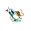
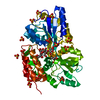
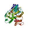
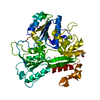
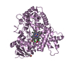
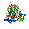
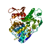
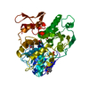
| Title | Revealing surface waters on an antifreeze protein by fusion protein crystallography | |||||||||
 Components Components | Fusion protein of Maltose-binding periplasmic protein and Type-3 ice-structuring protein HPLC 12 | |||||||||
 Keywords Keywords | ANTIFREEZE PROTEIN / fusion protein | |||||||||
| Function / homology |  Function and homology information Function and homology informationdetection of maltose stimulus / maltose transport complex / carbohydrate transport / carbohydrate transmembrane transporter activity / maltose binding / maltose transport / maltodextrin transmembrane transport / ATP-binding cassette (ABC) transporter complex, substrate-binding subunit-containing / ATP-binding cassette (ABC) transporter complex / cell chemotaxis ...detection of maltose stimulus / maltose transport complex / carbohydrate transport / carbohydrate transmembrane transporter activity / maltose binding / maltose transport / maltodextrin transmembrane transport / ATP-binding cassette (ABC) transporter complex, substrate-binding subunit-containing / ATP-binding cassette (ABC) transporter complex / cell chemotaxis / outer membrane-bounded periplasmic space / periplasmic space / DNA damage response / extracellular region / membrane Similarity search - Function | |||||||||
| Biological species |   Zoarces americanus (ocean pout) Zoarces americanus (ocean pout) | |||||||||
| Method |  X-RAY DIFFRACTION / SYNCHROTRON / MOLECULAR REPLACEMENT / Resolution: 1.94 Å X-RAY DIFFRACTION / SYNCHROTRON / MOLECULAR REPLACEMENT / Resolution: 1.94 Å | |||||||||
 Authors Authors | Sun, T. / Gauthier, S. / Campbell, R.L. / Davies, P.L. | |||||||||
 Citation Citation | Journal: J.Phys.Chem.B / Year: 2015 Title: Revealing Surface Waters on an Antifreeze Protein by Fusion Protein Crystallography Combined with Molecular Dynamic Simulations. Authors: Sun, T. / Gauthier, S.Y. / Campbell, R.L. / Davies, P.L. #1: Journal: J.Mol.Biol. / Year: 2001Title: Understanding the mechanism of ice binding by type III antifreeze proteins. Authors: Antson, A.A. / Smith, D.J. / Roper, D.I. / Lewis, S. / Caves, L.S. / Verma, C.S. / Buckley, S.L. / Lillford, P.J. / Hubbard, R.E. | |||||||||
| History |
|
- Structure visualization
| Structure viewer | Molecule: MolmilJmol/JSmol |
|---|
- Downloads & links
-Download
| PDBx/mmCIF format | 5c7r.cif.gz | 195.6 KB | Display | PDBx/mmCIF format |
|---|---|---|---|---|
| PDB format | pdb5c7r.ent.gz | 154.6 KB | Display | PDB format |
| PDBx/mmJSON format | 5c7r.json.gz | Tree view | PDBx/mmJSON format | |
| Others |  Other downloads Other downloads |
-Validation report
| Arichive directory | https://data.pdbj.org/pub/pdb/validation_reports/c7/5c7rftp://data.pdbj.org/pub/pdb/validation_reports/c7/5c7r | HTTPS FTP |
|---|
-Related structure data


-Links
 PDBj
PDBj


- Assembly
| Deposited unit | 
| ||||||||
|---|---|---|---|---|---|---|---|---|---|
| 1 | 
| ||||||||
| 2 | 
| ||||||||
| Unit cell |
|
-Components
| #1: Protein | Mass: 48512.152 Da / Num. of mol.: 2 Fragment: UNP P0AEY0 residues 27-384, UNP P19614 residues 1-63 Source method: isolated from a genetically manipulated source Source: (gene. exp.) Zoarces americanus (ocean pout)Gene: malE, Z5632, ECs5017 / Production host: References: UniProt: P0AEY0, UniProt: P19614, UniProt: P0AEX9*PLUS #2: Polysaccharide |   Source method: isolated from a genetically manipulated source Details: oligosaccharide / References: alpha-maltotriose #3: Chemical |   Mass: 96.063 Da / Num. of mol.: 2 / Source method: obtained synthetically / Formula: SO4 Mass: 96.063 Da / Num. of mol.: 2 / Source method: obtained synthetically / Formula: SO4#4: Water | ChemComp-HOH / |  Mass: 18.015 Da / Num. of mol.: 540 / Source method: isolated from a natural source / Formula: H2O Mass: 18.015 Da / Num. of mol.: 540 / Source method: isolated from a natural source / Formula: H2O |
|---|
-Experimental details
-Experiment
| Experiment | Method: X-RAY DIFFRACTION |
|---|
- Sample preparation
| Crystal | Density Matthews: 2.71 Å3/Da / Density % sol: 54.61 % |
|---|---|
| Crystal grow | Temperature: 298 K / Method: vapor diffusion, hanging drop / pH: 4.4 / Details: 1.75 M (NH4)2SO4, 100 mM NaOAC (pH 4.4) |
-Data collection
| Diffraction | Mean temperature: 100 K |
|---|---|
| Diffraction source | Source: SYNCHROTRON / Site: APS  / Beamline: 23-ID-D / Wavelength: 0.987 Å / Beamline: 23-ID-D / Wavelength: 0.987 Å |
| Detector | Type: PSI PILATUS 6M / Detector: PIXEL / Date: Aug 9, 2012 |
| Radiation | Protocol: SINGLE WAVELENGTH / Monochromatic (M) / Laue (L): M / Scattering type: x-ray |
| Radiation wavelength | Wavelength: 0.987 Å / Relative weight: 1 |
| Reflection | Resolution: 1.94→48.2 Å / Num. obs: 72126 / % possible obs: 96.7 % / Redundancy: 3.9 % / Rmerge(I) obs: 0.133 / Net I/σ(I): 9.36 |
| Reflection shell | Resolution: 1.94→2 Å / Redundancy: 3.4 % / Rmerge(I) obs: 1.362 / Mean I/σ(I) obs: 1.1 / % possible all: 86.2 |
- Processing
| Software |
| |||||||||||||||||||||||||||||||||||||||||||||||||||||||||||||||||||||||||||||||||||||||||||||||||||||||||
|---|---|---|---|---|---|---|---|---|---|---|---|---|---|---|---|---|---|---|---|---|---|---|---|---|---|---|---|---|---|---|---|---|---|---|---|---|---|---|---|---|---|---|---|---|---|---|---|---|---|---|---|---|---|---|---|---|---|---|---|---|---|---|---|---|---|---|---|---|---|---|---|---|---|---|---|---|---|---|---|---|---|---|---|---|---|---|---|---|---|---|---|---|---|---|---|---|---|---|---|---|---|---|---|---|---|---|
| Refinement | Method to determine structure: MOLECULAR REPLACEMENT Starting model: 1HG7,3G7W Resolution: 1.94→48.158 Å / SU ML: 0.25 / Cross valid method: FREE R-VALUE / σ(F): 1.99 / Phase error: 24.76 / Stereochemistry target values: ML
| |||||||||||||||||||||||||||||||||||||||||||||||||||||||||||||||||||||||||||||||||||||||||||||||||||||||||
| Solvent computation | Shrinkage radii: 0.9 Å / VDW probe radii: 1.11 Å / Solvent model: FLAT BULK SOLVENT MODEL | |||||||||||||||||||||||||||||||||||||||||||||||||||||||||||||||||||||||||||||||||||||||||||||||||||||||||
| Refinement step | Cycle: LAST / Resolution: 1.94→48.158 Å
| |||||||||||||||||||||||||||||||||||||||||||||||||||||||||||||||||||||||||||||||||||||||||||||||||||||||||
| Refine LS restraints |
| |||||||||||||||||||||||||||||||||||||||||||||||||||||||||||||||||||||||||||||||||||||||||||||||||||||||||
| LS refinement shell |
|
